. 2015 Dec 24:34:158.
doi: 10.1186/s13046-015-0277-8.

The number of polyploid giant cancer cells and epithelial-mesenchymal transition-related proteins are associated with invasion and metastasis in human breast cancer

Affiliations

The number of polyploid giant cancer cells and epithelial-mesenchymal transition-related proteins are associated with invasion and metastasis in human breast cancer

Fei Fei et al. J Exp Clin Cancer Res. .

Erratum in

Abstract

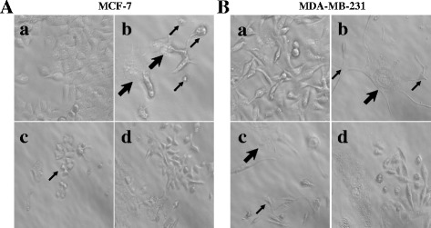
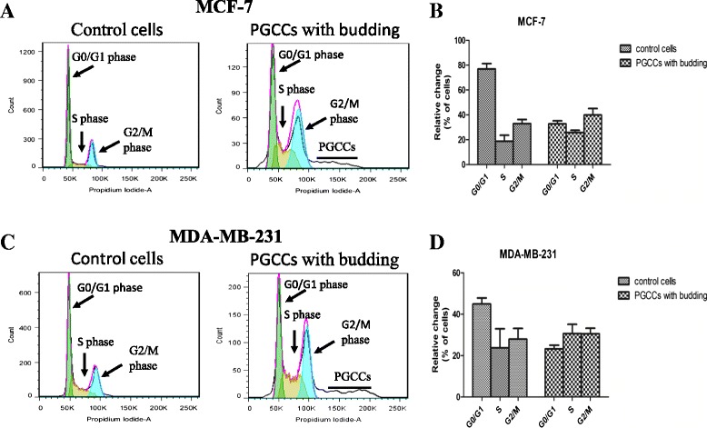
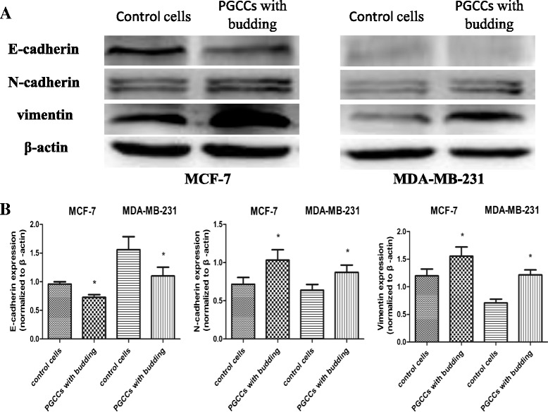
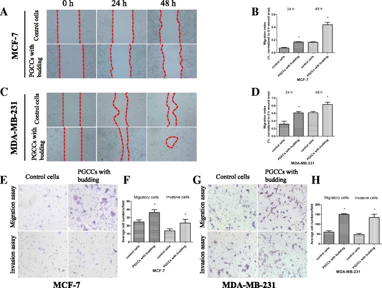
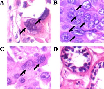
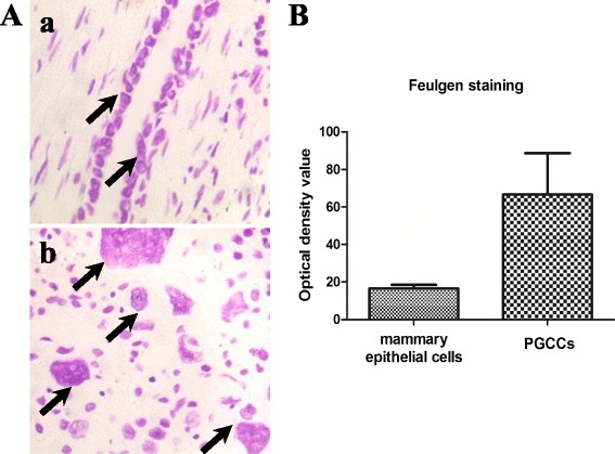
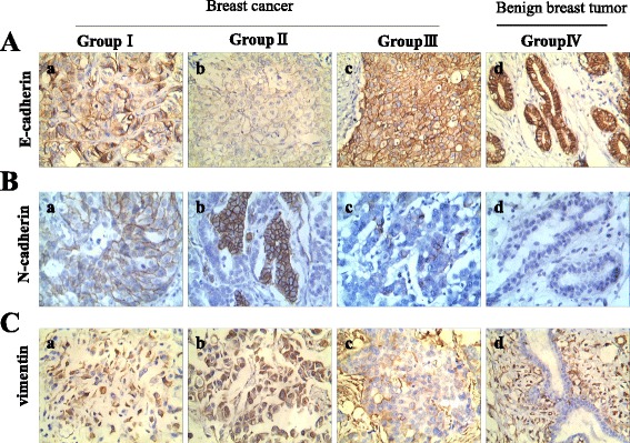
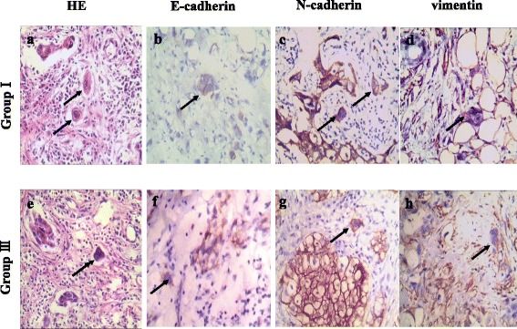
Background: Previously, we reported that polyploid giant cancer cells (PGCCs) induced by cobalt chloride (CoCl2) could have generated daughter cells with strong invasiveness and migration capabilities via asymmetric divisions. This study compared the expression of epithelial-mesenchymal transition (EMT)-related proteins, including E-cadherin, N-cadherin, and vimentin, between PGCCs and their daughter cells, and control breast cancer cell lines MCF-7 and MDA-MB-231. The clinicopathological significance of EMT-related protein expression in human breast cancer was analyzed.

Methods: Western blot was used to compare the expression levels of E-cadherin, N-cadherin, and vimentin in breast cancer lines MCF-7 and MDA-MB-231, between PGCCs with budding daughter cells and control breast cancer cells. Furthermore, 167 paraffin-embedded breast tumor tissue samples were analyzed, including samples obtained from 52 patients with primary breast cancer with lymph node metastasis (group I) and their corresponding lymph node metastatic tumors (group II), 52 patients with primary breast cancer without metastasis (group III), and 11 patients with benign breast lesions (group IV). The number of PGCCs was compared among these four groups.

Results: The number of PGCCs increased with the malignant grade of breast tumor. Group IIhad the highest number of PGCCs and the differences among group I, II, III and IV had statistically significance (P =0.000). In addition, the expression of E-cadherin (P = 0.000), N-cadherin (P = 0.000), and vimentin (P = 0.000) was significantly different among the four groups. Group II exhibited the highest expression levels of N-cadherin and vimentin and the lowest expression levels of E-cadherin.

Conclusions: These data suggest that the number of PGCCs and the EMT-related proteins E-cadherin, N-cadherin, and vimentin may be valuable biomarkers to assess metastasis in patients with breast cancer.


Figures

Fig. 1
Fig. 1
PGCCs with budding daughter cells in MCF-7 and MDA-MB-231 cells. A. MCF-7 PGCCs and control MCF-7 cells. a. Control MCF-7 cells (×400). b. MCF-7 PGCCs induced by 450 μM CoCl2 treatment for 72 h (×400). Small black arrowheads indicate budded daughter cells; large black arrow heads indicate PGCCs. c. PGCCs generated daughter cells via budding 10–15 days after CoCl2 treatment. Black arrowheads indicate budded daughter cells (×100). d. Fast reproduction of PGCCs by generated daughter cells via budding (×100). B. MDA-MB-231 PGCCs and control MDA-MB-231 cells. a. Control MDA-MB-231 cells (×400). b. MDA-MB-231 PGCCs induced by 300 μM CoCl2 treatment for 72 h (×400). Small black arrowheads indicate budded daughter cells; large black arrowheads indicate PGCCs. c. PGCCs generated daughter cells via budding 10–15 days after CoCl2 treatment. Small black arrowheads indicate budded daughter cells; large black arrowheads indicate PGCCs (×100). d. Recovery of PGCCs by generated budding daughter cells (×100)
Fig. 2
Fig. 2
Alteration of cell cycle in breast cancer cell lines after CoCl2 treatment. Clear alteration of the cell cycle as analyzed by flow cytometry was observed in MCF-7 (a) and MDA-MB-231 (c) cells treated with CoCl2. The bar graphs in (b) and (d) depict the relative changes to cell percentage in different phases of the cell cycle in PGCCs with budding compared to that of control cells. All data represent the mean ± SD of three independent cultures
Fig. 3
Fig. 3
E-cadherin, N-cadherin, and vimentin expression in PGCCs with budding and control MCF-7 and MDA-MB-231 cells. a Western blot was used to detect differences in E-cadherin, N-cadherin, and vimentin expression in MCF-7 and MDA-MB-231 cells before and after CoCl2 treatment. b Quantitative results of protein expression differences are shown as histograms. The corresponding densitometric analyses of the protein bands performed using Image-J software were normalized to the signal of β-actin. Each bar represents the mean ± SD of three independent experiments (*p < 0.05)
Fig. 4
Fig. 4
CoCl2 increases the migration and invasion of breast cancer cells. a Representative images of the wound-healing assay for MCF-7 cells at different times (×40). b MCF-7 cell migration is shown as a wound-healing index quantified by measuring at least three different wound areas. c Representative images of the wound-healing assay for MDA-MB-231 cells at different times (×40). d Quantitative data of MDA-MB-231 cell migration between control cells and PGCCs with budding. e, g Transwell migration and invasion assays were performed in control MCF-7 and MDA-MB-231 cells and PGCCs with budding (×100). Upper panels indicate the migration and lower panels show cell invasion. f, h Quantitative results of transwell migration and invasion assay in MCF-7 and MDA-MB-231 cells
Fig. 5
Fig. 5
PGCCs in human breast tumors. a PGCCs in primary breast cancer with metastasis (group I; black arrowheads, H&E, ×400). b PGCCs in metastatic tumors (group II; black arrowheads, H&E, ×400). c PGCCs in primary breast cancer without metastasis (group III; black arrowheads, H&E, ×400). d PGCCs are absent in benign breast tumors (group IV; H&E, ×400)
Fig. 6
Fig. 6
Feulgen staining and MOD determination of PGCCs and control mammary epithelial cells. A. Feulgen staining between PGCCs and control mammary epithelial cells. a. Feulgen staining of mammary epithelial cells (black arrowheads, ×200). b. Feulgen staining of PGCCs in breast cancer (black arrowheads, ×200). B. Quantitative results of MOD determination in PGCCs and control mammary epithelial cells
Fig. 7
Fig. 7
Expression of E-cadherin, N-cadherin, and vimentin in human breast tumor tissues. A. E-cadherin expression in (a) primary breast cancer with lymph node metastasis (group I), (b) corresponding metastatic cancer (group II), (c) primary breast cancer without metastasis (group III), and (d) benign breast tumor (group IV) (×200). B. N-cadherin expression in (a) primary breast cancer with lymph node metastasis (group I), (b) corresponding metastatic tumor (group II), (c) primary breast cancer without metastasis (group III), and (d) benign breast tumor (group IV) (×200). C. Vimentin expression in (a) primary breast cancer with lymph node metastasis (group I), (b) corresponding metastatic tumor (group II), (c) primary breast cancer without metastasis (group III), and (d) benign breast tumor (group IV) (×200)
Fig. 8
Fig. 8
Expression of E-cadherin, N-cadherin, and vimentin in single stroma PGCCs of human breast cancer. a Single stroma PGCC in human breast cancer with lymph node metastasis (Black arrow head, H&E, ×200). b E-cadherin, (c) N-cadherin, (d) vimentin expression of single stroma PGCC in human breast cancer with lymph node metastasis (Black arrow head, IHC,×200). e Single stroma PGCC in human breast cancer without metastasis (Black arrow head, H&E, ×200). b E-cadherin, (c) N-cadherin, (d) vimentin expression of single stroma PGCC in human breast cancer without metastasis (Black arrow head, IHC, ×200)

References

    1. Torre LA, Bray F, Siegel RL, Ferlay J, Lortet-Tieulent J, Jemal A. Global cancer statistics, 2012. CA Cancer J Clin. 2015;65(2):87–108. doi: 10.3322/caac.21262. - DOI - PubMed
    1. Kennecke H, Yerushalmi R, Woods R, Cheang MC, Voduc D, Speers CH, et al. Metastatic behavior of breast cancer subtypes. J. Clin. Oncol. Off. J. Am. Soc. Clin. Oncol. 2010;28(20):3271–7. doi: 10.1200/JCO.2009.25.9820. - DOI - PubMed
    1. Zhang S, Mercado-Uribe I, Xing Z, Sun B, Kuang J, Liu J. Generation of cancer stem-like cells through the formation of polyploid giant cancer cells. Oncogene. 2014;33(1):116–28. doi: 10.1038/onc.2013.96. - DOI - PMC - PubMed
    1. Zhang S, Mercado-Uribe I, Liu J. Generation of erythroid cells from fibroblasts and cancer cells in vitro and in vivo. Cancer Lett. 2013;333(2):205–12. doi: 10.1016/j.canlet.2013.01.037. - DOI - PMC - PubMed
    1. Marusyk A, Almendro V, Polyak K. Intra-tumour heterogeneity: a looking glass for cancer? Nat Rev Cancer. 2012;12(5):323–34. doi: 10.1038/nrc3261. - DOI - PubMed

Publication types

MeSH terms