. 2015 Dec 21;35(6):698-712.
doi: 10.1016/j.devcel.2015.11.022.

The E3 Ubiquitin Ligase TRIM9 Is a Filopodia Off Switch Required for Netrin-Dependent Axon Guidance

Affiliations

The E3 Ubiquitin Ligase TRIM9 Is a Filopodia Off Switch Required for Netrin-Dependent Axon Guidance

Shalini Menon et al. Dev Cell. .

Abstract

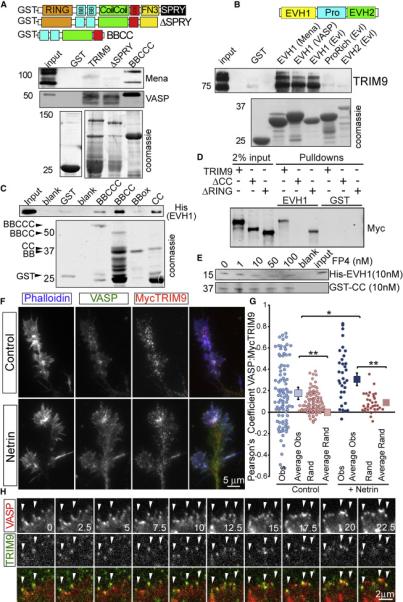
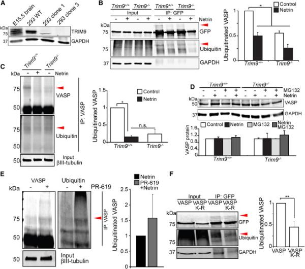
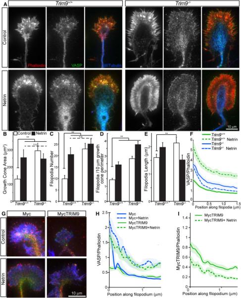
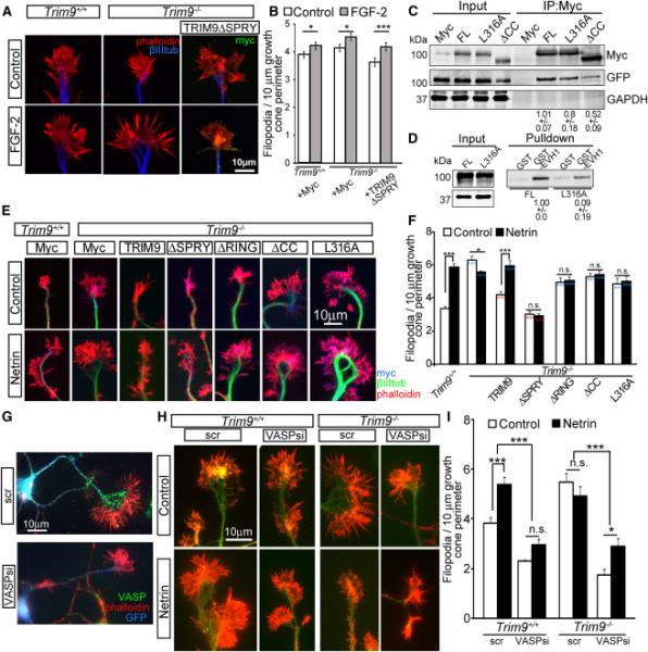
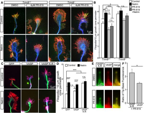
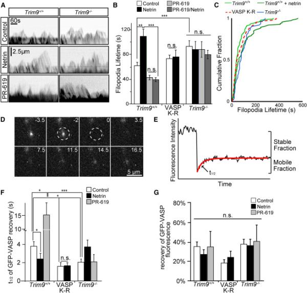
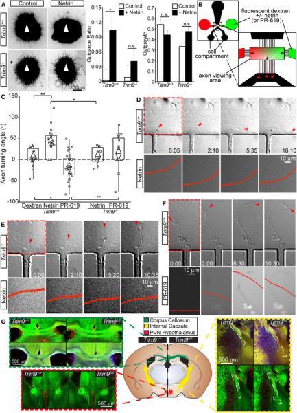
Neuronal growth cone filopodia contain guidance receptors and contribute to axon guidance; however, the mechanism by which the guidance cue netrin increases filopodia density is unknown. Here, we demonstrate that TRIM9, an E3 ubiquitin ligase that localizes to filopodia tips and binds the netrin receptor DCC, interacts with and ubiquitinates the barbed-end polymerase VASP to modulate filopodial stability during netrin-dependent axon guidance. Studies with murine Trim9(+/+) and Trim9(-/-) cortical neurons, along with a non-ubiquitinatable VASP mutant, demonstrate that TRIM9-mediated ubiquitination of VASP reduces VASP filopodial tip localization, VASP dynamics at tips, and filopodial stability. Upon netrin treatment, VASP is deubiquitinated, which promotes VASP tip localization and filopodial stability. Trim9 deletion induces axon guidance defects in vitro and in vivo, whereas a gradient of deubiquitinase inhibition promotes axon turning in vitro. We conclude that a gradient of TRIM9-mediated ubiquitination of VASP creates a filopodial stability gradient during axon turning.


Figures

Figure 1
Figure 1. TRIM9 is a brain-enriched Ena/VASP interaction partner
A-B) Binding assays with purified GST fusion proteins incubated in E15.5 brain lysate. Coomassie stained gels of recombinant proteins shown in lower panels. A) GST-BBox-coiled-coil-COS (BBCCC) of TRIM9 precipitates endogenous Mena and VASP. B) The GST-EVH1 domain of Mena, VASP and EVL interacts with endogenous TRIM9. C) In vitro binding assay showing that GST-Coiled Coil (CC) domain of TRIM9 directly binds His-EVH1 domain of VASP. D) Binding assay showing that GST-EVH1 precipitates Myc-TRIM9 and Myc-TRIM9ΔRING from cell lysate, but not Myc-TRIM9ΔCC. E) A 10-fold excess of FP4 containing peptide does not block the CC-EVH1 interaction. F) Axonal growth cones of control and netrin-treated cortical neurons stained for VASP (green), MycTRIM9 (red) and phalloidin (blue). G) Quantification of Pearson's correlation coefficient within filopodia (Obs=Observed measurements, Rand=pixels from one image randomized). Squares represent means +/− 95% CI. H) Montage of TIRF images of GFP-VASP (red) and mCherry-TRIM9 (green). Arrowheads denote filopodia tips in which TRIM9 and VASP colocalize, time in seconds. (See also Movies S1-3, FigS1)
Figure 2
Figure 2. VASP ubiquitination occurs in the presence of TRIM9 and is lost following netrin treatment
A) Immunoblot for TRIM9 and GAPDH in E15.5 brain lysates, HEK293 lysates: TRIM9+/+ (WT), TRIM9+/− (clone 1) and TRIM9−/− (clone 3). B) Input and GFP-VASP immunoprecipitation (IP) from ubiquitination assay immunoblotted (IB) for GFP, ubiquitin and GAPDH. Shown are GFP-VASP (75kDa) in TRIM9+/+ and TRIM9−/− cell lysates and ~25kDa heavier VASP band present in TRIM9+/+ lysate that co-migrates with ubiquitin (red arrowheads). Plot shows quantification of VASP ubiquitination. C) Endogenous VASP ubiquitination in TRIM9+/+ and TRIM9−/− neurons at 2DIV. A higher molecular weight VASP+ band (red arrowhead) that co-migrates with ubiquitin is seen in TRIM9+/+ cortical neurons. D) Endogenous VASP in TRIM9+/+ and TRIM9−/− control neurons or neurons treated with MG132 and/or 600 ng/ml netrin. IB for VASP (50kDa) and GAPDH. No difference in VASP protein levels detected between genotypes or treatment conditions. E) Endogenous VASP ubiquitination in TRIM9+/+ cortical neurons treated with netrin or netrin and 4 μM PR-619. Elevated ubiquitination is observed upon PR-619 treatment. F) Ubiquitination assay and quantification of wildtype VASP and VASP K-R mutant. The VASP K-R mutant exhibits a reduction in the ~25kDa heavier VASP band that co-migrates with ubiquitin (red arrowheads). All error bars represent SEM. (See also FigS2)
Figure 3
Figure 3. Deletion of TRIM9 increases growth cone size and filopodia density and alters VASP localization to filopodia tips
A-F) Images and quantification of axonal growth cones from control and netrin-treated TRIM9+/+ and TRIM9−/− neurons, stained for phalloidin (red, left), VASP (green, middle) and βIII tubulin (blue, merge). Quantification of B) growth cone area +/−SEM, C) growth cone filopodia number +/−SEM, D) density of growth cone filopodia +/−SEM, E) filopodia length +/−SEM, and F) VASP fluorescence intensity relative to phalloidin +/− 95% CI from the tip of filopodia into growth cone. G-I) Images and quantification of TRIM9−/− growth cones stained for VASP (red), phalloidin (blue), and Myc (green, Myc or MycTRIM9). H) VASP fluorescence intensity normalized to phalloidin +/− 95% CI from filopodia tip into growth cone. Expression of TRIM9 rescues VASP localization. I) MycTRIM9 fluorescence intensity normalized to phalloidin +/− 95% CI from filopodia tip into growth cone. (See also FigS3)
Figure 4
Figure 4. TRIM9 constrains filopodia density through VASP
A-B) Images and quantification of filopodia +/−SEM in axonal growth cones from control and FGF2-treated TRIM9+/+ and TRIM9−/− cortical neurons expressing Myc or MycTRIM9ΔSPRY, stained for Myc (green), βIII tubulin (blue) and phalloidin (red). C) TRIM9 dimerization assay IB for GFP-TRIM9 (IB:GFP) co-IP with Myc-TRIM9 variants (IP:Myc, IB:Myc). GAPDH is loading control. Numbers indicate levels of coIP GFP-TRIM9 +/−SEM. D) Binding assay immunoblot showing MycTRIM9 and TRIM9-L316A interaction with GST-EVH1. Numbers denote relative levels of MycTRIM9 variant precipitated by GST-EVH1 beads +/−SEM. E-F) Images and quantification of filopodia +/−SEM in axonal growth cones from control and netrin-treated TRIM9+/+ and TRIM9−/− neurons stained for Myc (blue: Myc, Myc-TRIM9 or TRIM9 mutants), βIII tubulin (green) and phalloidin (red). *denotes significance compared to TRIM9+/+ and *denotes significance compared to TRIM9−/−. G) Representative images of TRIM9−/− neurons transfected with scrambled (scr) or VASP siRNA, along with GFP to identify transfected cells. GFP (blue), VASP immunostaining (green), phalloiding (red). H-I) Images and quantification of axonal growth cone filopodia +/−SEM from control and netrin-treated TRIM9+/+ and TRIM9−/− cortical neurons transfected with scramble (scr) or VASP siRNA (VASPsi), stained for GFP (green) and phalloidin (red). (See also FigS4)
Figure 5
Figure 5. VASP deubiquitination is required for netrin-dependent increases in filopodia density
A-B) Images and quantification of filopodia +/−SEM in axonal growth cones from control, PR-619 netrin or PR-619/netrin treated TRIM9+/+ and TRIM9−/− neurons, stained for VASP (green), βIII tubulin (blue) and phalloidin (red). C-D) Images and quantification of filopodia +/−SEM in TRIM9+/+ growth cones expressing GFP, GFP-VASP or GFP-VASP K-R, stained for GFP (blue), βIII tubulin (green) and phalloidin (red). E) TRIM9+/+ filopodia containing GFP-VASP K-R (green) and mCherry-VASP (red) before and after PR-619 treatment. F) Ratio of fluorescence intensity at filopodial tip:filopodial base of GFP-VASP K-R or mCherry-VASP +/−SEM in PR-619 treated TRIM9+/+ neurons. (See also FigS5)
Figure 6
Figure 6. Ubiquitination of VASP reduces filopodia lifetime and the rate of VASP dissociation from filopodia tips
A) Example kymographs of axonal growth cone filopodia from TRIM9+/+ and TRIM9−/− neurons expressing mCherry. B) Filopodial lifetimes +/−SEM in control, netrin, and PR-619 treated TRIM9+/+ and TRIM9−/− neurons and TRIM9+/+ neurons expressing GFP-VASP K-R. C) Cumulative fraction plot of filopodial lifetime demonstrating intermediate phenotype of VASP K-R expressing neurons. D) Image montage of GFP-VASP FRAP at a filopodium tip. Bleach denoted by dashed region, time before and after bleaching in seconds. E) Example of fluorescence intensity data fit to a single exponential (red line), depicting percent fluorescence recovery and t1/2 of fluorescence recovery. F) Fluorescence recovery halftime (t1/2) +/−SEM and G) mean % fluorescence recovery +/−SEM for indicated conditions. (See also Movies S4, S5, FigS6). VASP K-R was expressed in TRIM9+/+ neurons. (See also FigS6)
Figure 7
Figure 7. Deletion of TRIM9 disrupts axon guidance
A) TRIM9+/+ but not TRIM9−/− cortical explants exhibit biased neurite outgrowth (βIII tubulin staining) towards netrin (arrowhead). B) Schematic representation of a micropass gradient device with zoomed view of the axon viewing area depicting the dextran gradient and axon growth into the viewing area, arrows indicate direction of fluid flow. C) Turning angles for TRIM9+/+ axons in a dextran gradient, dextran/netrin or dextran/PR-619 gradient and TRIM9−/− axons in a dextran/netrin gradient or dextran/PR-619 gradient. D) DIC images (top) showing a TRIM9+/+ axon turning towards a higher netrin concentration, as seen by the gradient in epifluorescence images (bottom). The red arrowhead denotes the front of the growth cone, the red box denotes the area for which epifluorescence is shown. Time in hours:minutes. Quantification of dextran fluorescence intensity across the region within each epifluorescence image (red), x and y axes are kept constant to demonstrate the stability of the gradient over time. E) TRIM9−/− axon (arrowhead) failing to turn toward the higher netrin concentrations, gradient displayed as in D. F) TRIM9+/+ axon (arrowhead) turning down a PR-619 gradient, displayed as in D. G) Regions of interest (ROI) from coronal sections of Nex-Cre/TauloxP-stop-loxPGFP/TRIM9fl/fl and TRIM9+/+ littermates reveal aberrant cortical axon projections patterns in the corpus callosum (green dashed box), the paraventricular hypothalamic nuclei (red), and the internal capsule (yellow). Green is GFP, red and blue demarcate GFAP in separate littermate pairs. The locations of ROIs and associated defects are denoted in the coronal schematic, n=3 littermate pairs. (See also Movies S6-7, FigS7)

Comment in

References

    1. Altun M, Kramer HB, Willems LI, McDermott JL, Leach CA, Goldenberg SJ, Kumar KGS, Konietzny R, Fischer R, Kogan E, et al. Activity-based chemical proteomics accelerates inhibitor development for deubiquitylating enzymes. Chem. Biol. 2011;18:1401–1412. - PubMed
    1. Applewhite DA, Barzik M, Kojima S-I, Svitkina TM, Gertler FB, Borisy GG. Ena/VASP proteins have an anti-capping independent function in filopodia formation. Mol Biol Cell. 2007;18:2579–2591. - PMC - PubMed
    1. Barzik M, Kotova TI, Higgs HN, Hazelwood L, Hanein D, Gertler FB, Schafer DA. Ena/VASP proteins enhance actin polymerization in the presence of barbed end capping proteins. J Biol Chem. 2005;280:28653–28662. - PMC - PubMed
    1. Bear JE, Svitkina TM, Krause M, Schafer DA, Loureiro JJ, Strasser GA, Maly IV, Chaga OY, Cooper JA, Borisy GG, et al. Antagonism between Ena/VASP proteins and actin filament capping regulates fibroblast motility. Cell. 2002;109:509–521. - PubMed
    1. Boëda B, Knowles PP, Briggs DC, Murray-Rust J, Soriano E, Garvalov BK, McDonald NQ, Way M. Molecular recognition of the Tes LIM2-3 domains by the actin-related protein Arp7A. J Biol Chem. 2011;286:11543–11554. - PMC - PubMed

Publication types

LinkOut - more resources