. 2015 Dec 24:15:1009.
doi: 10.1186/s12885-015-2025-z.

Role of HLA-G and extracellular vesicles in renal cancer stem cell-induced inhibition of dendritic cell differentiation

Affiliations

Role of HLA-G and extracellular vesicles in renal cancer stem cell-induced inhibition of dendritic cell differentiation

Cristina Grange et al. BMC Cancer. .

Abstract

Background: Tumor immune-escape has been related to the ability of cancer cells to inhibit T cell activation and dendritic cell (DC) differentiation. We previously identified a tumor initiating population, expressing the mesenchymal marker CD105, which fulfills the criteria for definition as cancer stem cells (CD105(+) CSCs) able to release extracellular vesicles (EVs) that favor tumor progression and metastases. The aim of the present study was to compare the ability of renal CSCs and derived EVs to modulate the behavior of monocyte-derived DCs with a non-tumor initiating renal cancer cell population (CD105(-) TCs) and their EVs.

Methods: Maturation of monocyte-derived DCs was studied in presence of CD105(+) CSCs and CD105(-) TCs and their derived EVs. DC differentiation experiments were evaluated by cytofluorimetric analysis. T cell proliferation and ELISA assays were performed. Monocytes and T cells were purified from peripheral blood mononuclear cells obtained from healthy donors.

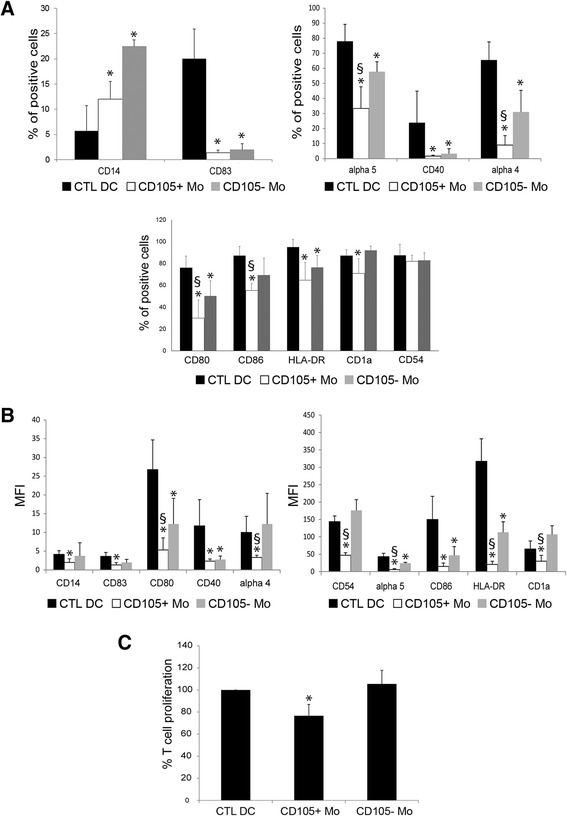
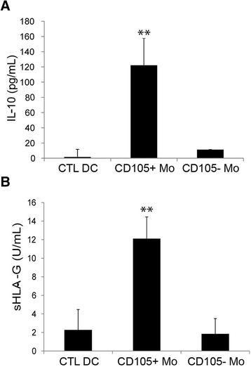
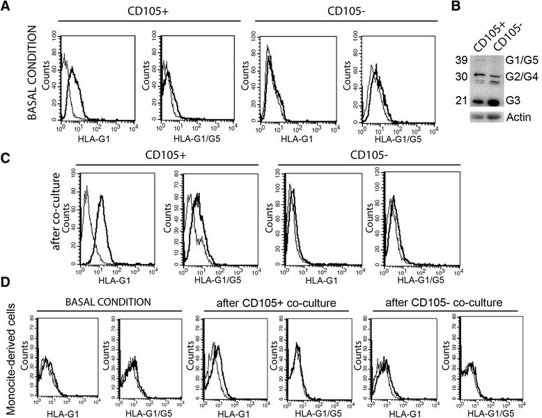
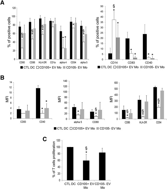
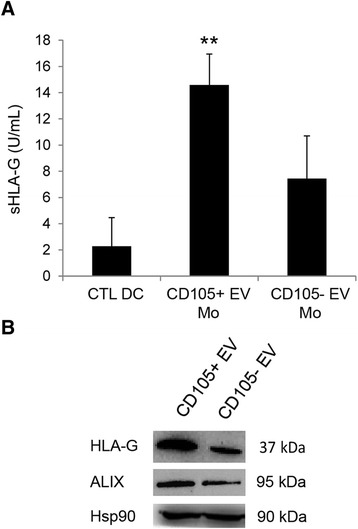
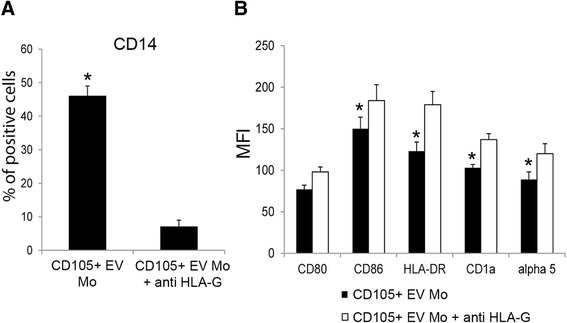
Results: The results obtained demonstrate that both CD105(+) CSCs and CD105(-) TCs impaired the differentiation process of DCs from monocytes. However, the immune-modulatory effect of CD105(+) CSCs was significantly greater than that of CD105(-) TCs. EVs derived from CD105(+) CSCs and in less extent, those derived from CD105(-) TCs retained the ability to impair monocyte maturation and T cell activation. The mechanism has been mainly related to the expression of HLA-G by tumor cells and to its release in a form associated to EVs. HLA-G blockade significantly reduced the inhibitory effect of EVs on DC differentiation.

Conclusions: In conclusion, the results of the present study indicate that renal cancer cells and in particular CSCs and derived EVs impair maturation of DCs and T cell immune response by a mechanism involving HLA-G.


Figures

Fig. 1
Fig. 1
Renal cancer cells suppressed monocyte-derived DC differentiation and their ability to stimulate T cell proliferation. a Mean percentage expression ± SD of CD14, CD83, α5 integrin, CD40, α4 integrin, CD80, CD86, HLA-DR, CD1a and CD54 by monocyte-derived DCs differentiated in the presence or in absence (CTL DC) of CD105+ CSCs (CD105+ Mo) or CD105- TCs (CD105- Mo). Results were obtained from 6 independent experiments. ANOVA with Newman Keuls multicomparison test was performed: *p < 0.05 CD105+ Mo and CD105- Mo versus CTL DC; § p < 0.05 CD105+ Mo versus CD105- Mo. b MFI ± SD of CD14, CD83, CD80, CD40, α4 integrin, CD54, α5 integrin, CD86, HLA-DR and CD1a of monocyte-derived DCs differentiated in the presence or in absence (CTL DC) of CD105+ CSCs (CD105+ Mo) or CD105- TCs (CD105- Mo). Results were obtained from 6 independent experiments. ANOVA with Newman Keuls multicomparison test was performed: *p < 0.05 CD105+ Mo and CD105- Mo versus CTL DC; § p < 0.05 CD105+ Mo versus CD105- Mo. c Monocyte-derived DCs differentiated in the presence or in absence (CTL DC) of CD105+ CSCs (CD105+ Mo) or CD105- TCs (CD105- Mo) were plated at cell concentration of 2x10⁴ with 1x10⁵ T CD3+ lymphocytes. Forty eight hours later T-cell proliferation was assessed. Data are expressed as mean ± SD of percent variation of T-cell proliferation in the presence of DCs differentiated in presence of renal cancer cells in respect to T-cell proliferation in presence of DCs matured in the absence of cells (established as 100 %). Results were obtained from 5 independent experiments. ANOVA with Newman Keuls multicomparison test was performed: *p < 0.05 CD105+ Mo versus all the other conditions
Fig. 2
Fig. 2
Co-culture of monocyte-derived cells with CD105+ CSCs induced the release of IL-10 and sHLA-G. Cell supernatants were harvested to detect IL-10 (a) and sHLA-G (b) production by ELISA, after 7 days of co-culture of monocyte-derived cells in the presence or absence (CTL DC) of renal cancer cells (CD105+ CSCs and CD105- TCs). Results were obtained from 3 independent experiments and expressed as mean ± SD. ANOVA with Newman Keuls multicomparison test was performed: ** p < 0.001 CD105+ CSC Mo versus all the other conditions
Fig. 3
Fig. 3
HLA-G expression and up-regulation by CD105+ CSCs. a Representative cytofluorimetric analysis of HLA-G expression on CD105+ CSCs and CD105- TCs membrane (G1) and intra-cytoplasmic (G1/G5) staining in basal culture condition (n = 4). b Western Blot analyses confirmed the presence of several isoforms of HLA-G in CD105+ CSCs and CD105- TCs. c Representative cytofluorimetric analysis of membrane (G1) and intra-cytoplasmic (G1/G5) staining of HLA-G on CD105+ CSCs and CD105- TCs after 7 days of co-culture with monocyte-derived cells (n = 4). d Representative cytofluorimetric analysis of HLA-G expression on monocyte-derived cell membrane (G1) and intra-cytoplasmic (G1/G5) in basal condition and after co-culture with CD105+ CSCs and CD105- TCs (n = 4)
Fig. 4
Fig. 4
EVs shed by renal cancer cells inhibited monocyte-derived DC differentiation and their ability to stimulate T cell proliferation. a Mean percentage expression ± SD of CD80, CD86, HLA-DR, CD1a, α4 integrin, CD54, α5 integrin, CD14, CD83 and CD40 by monocyte-derived DCs differentiated in the presence or in absence (CTL DC) of CD105+ EVs (CD105+ EV Mo) or CD105- EVs (CD105- EV Mo). Results were obtained from 6 independent experiments. ANOVA with Newman Keuls multicomparison test was performed: *p < 0.05 CD105+ EV Mo and CD105- EV Mo versus CTL DC; § p < 0.05 CD105+ EV Mo versus CD105- EV Mo. b MFI ± SD of CD83, CD40, α5 integrin CD80, CD86, HLA-DR and CD54 of monocyte-derived DCs differentiated in the presence or in absence (CTL DC) of CD105+ EVs (CD105+ EV Mo) or CD105- EVs (CD105- EV Mo). Results were obtained from 6 independent experiments. ANOVA with Newman Keuls multicomparison test was performed: *p < 0.05 CD105+ EV Mo and CD105- EV Mo versus CTL DC; § p < 0.05 CD105+ EV Mo versus CD105- EV Mo. c Monocyte-derived DCs differentiated in the presence or in absence (CTL DC) of CD105+ EVs (CD105+ EV Mo) or CD105- EVs (CD105- EV Mo) were plated at cell concentration of 2x10⁴ with 1x10⁵ T CD3+ lymphocytes. Forty eight hours later T-cell proliferation was assessed. Data are expressed as mean ± SD of percent variation of T-cell proliferation in the presence of DCs differentiated in the presence of renal cancer cells in respect to T-cell proliferation in presence of DCs matured in the absence of EVs (established as 100 %). Results were obtained from 4 independent experiments. ANOVA with Newman Keuls multicomparison test was performed: *p < 0.05 CD105+ EV Mo versus all the other conditions
Fig. 5
Fig. 5
Treatment of monocyte-derived cells with CD105+ EVs induced a release of sHLA-G. a Supernatants were harvested to detect sHLA-G production by ELISA, after 7 days of culture of monocyte-derived cells stimulated with EVs shed by renal cancer cells (CD105+ CSCs and CD105- TCs). Results were obtained from 3 independent experiments and expressed as mean ± SD. ANOVA with Newman Keuls multicomparison test was performed: ** p < 0.001 CD105+ EV Mo versus all the other conditions. b Representative Western Blot analysis showing the presence of HLA-G and Alix within EVs. Hsp90 was used as normalization. Four experiments were performed with similar results
Fig. 6
Fig. 6
The use of specific blocking antibody against HLA-G partially reverted the inhibitory effect on monocyte-derived DC differentiation induced by CD105+ CSC EVs. a Mean percentages ± SD of CD14 expression by monocyte-derived DCs stimulated with CD105+ EVs in the presence or in absence (CD105+ EV Mo) of specific blocking antibody against HLA-G (CD105+ EV Mo + anti HLA-G). Results were obtained from 2 independent experiments. T Student test was performed: *p < 0.05 CD105+ EV Mo versus CD105+ EV Mo + anti HLA-G. b MFI ± SD of CD80, CD86, ΗLA-DR, CD1a and α5 integrin of monocyte-derived DCs stimulated with CD105+ EVs in the presence or in absence (CD105+ EV Mo) of specific blocking antibody against HLA-G (CD105+ EV Mo + anti HLA-G). T Student test was performed: *p < 0.05 CD105+ EV Mo versus CD105+ EV Mo + anti HLA-G

Similar articles

Cited by

References

    1. Zins K, Sioud M, Aharinejad S, Lucas T, Abraham D. Modulating the tumor microenvironment with RNA interference as a cancer treatment strategy. Methods Mol Biol. 2015;1218:143–161. doi: 10.1007/978-1-4939-1538-5_9. - DOI - PubMed
    1. Bussolati B, Bruno S, Grange C, Ferrando U, Camussi G. Identification of a tumor-initiating stem cell population in human renal carcinomas. FASEB J. 2008;22:3696–3705. doi: 10.1096/fj.08-102590. - DOI - PubMed
    1. Grange C, Tapparo M, Collino F, Vitillo L, Damasco C, Deregibus MC, et al. Microvesicles released from human renal cancer stem cells stimulate angiogenesis and formation of lung premetastatic niche. Cancer Res. 2011;71:5346–5356. doi: 10.1158/0008-5472.CAN-11-0241. - DOI - PubMed
    1. Muralidharan-Chari V, Clancy JW, Sedgwick A, D'Souza-Schorey C. Microvesicles: mediators of extracellular communication during cancer progression. J Cell Sci. 2010;123:1603–1611. doi: 10.1242/jcs.064386. - DOI - PMC - PubMed
    1. Van Doormaal FF, Kleinjan A, Di Nisio M, Büller HR, Nieuwland R. Cell-derived microvesicles and cancer. Neth J Med. 2009;67:266–273. - PubMed

Publication types

MeSH terms