. 2016 May:146:43-53.
doi: 10.1016/j.exer.2015.12.006. Epub 2015 Dec 17.

Three-dimensional arrangement of elastic fibers in the human corneal stroma

Affiliations

Three-dimensional arrangement of elastic fibers in the human corneal stroma

Philip N Lewis et al. Exp Eye Res. 2016 May.

Abstract

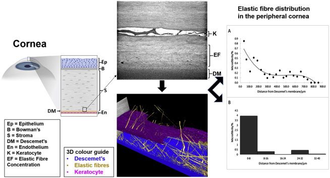
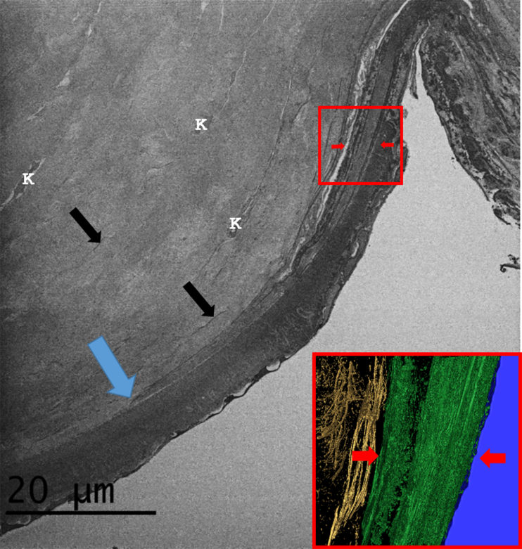
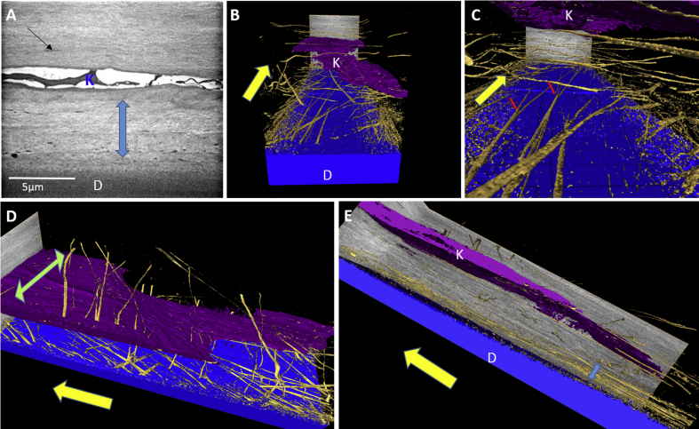
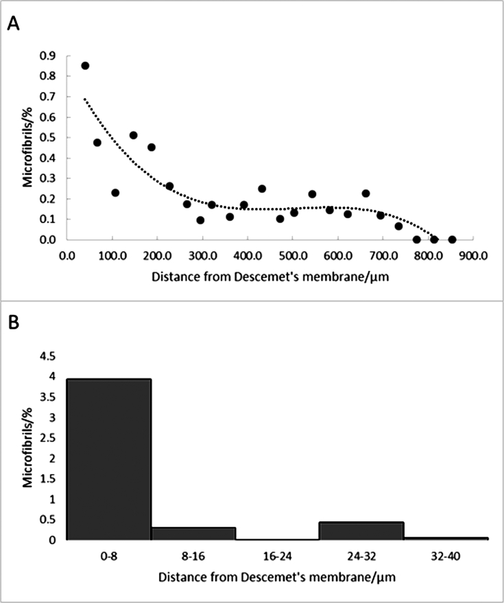
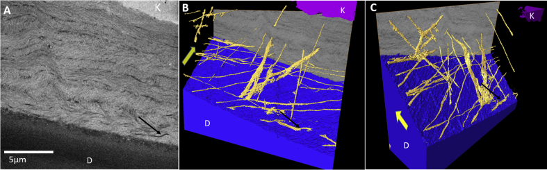
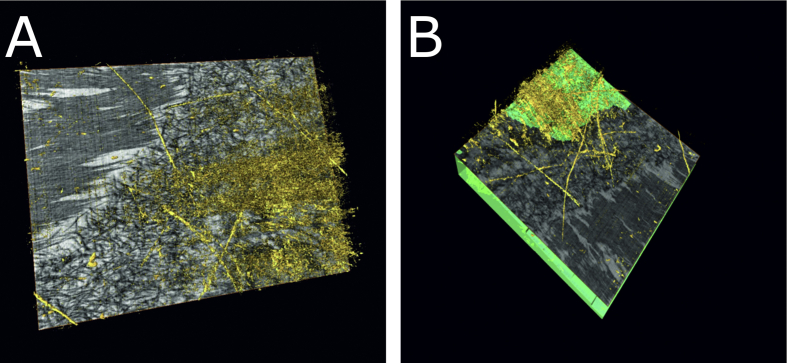
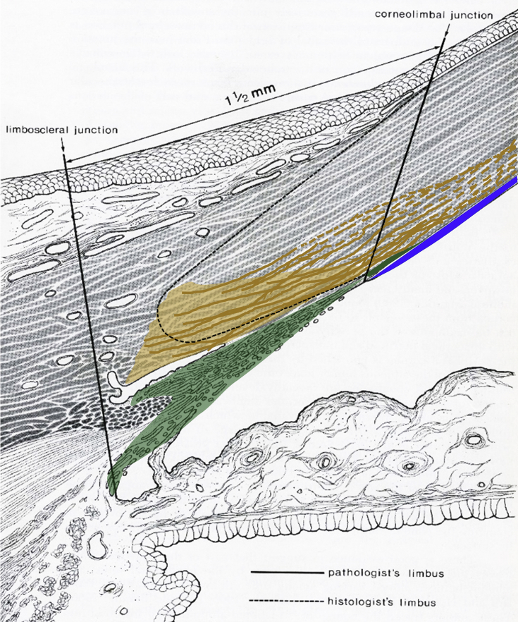
The cornea is the main refracting lens in the eye. As part of the outer tunic it has to be resilient, a property conferred by the organisation of the constituent collagen. It also has to be sufficiently elastic to regain its exact shape when deformed, in order not to distort the retinal image. The basis of this elasticity is not fully understood. The purpose of this study was to characterise in three dimensions the arrangement and distribution of elastic fibers in the human corneal stroma, using serial block face scanning electron microscopy. We have demonstrated that there exists a complex network of elastic fibers that appear to originate in the sclera or limbus. These appear as elastic sheets in the limbus and peripheral cornea immediately above the trabecular meshwork which itself appears to extend above Descemet's membrane in the peripheral stroma. From these sheets, elastic fibers extend into the cornea; moving centrally they bifurcate and trifurcate into narrower fibers and are concentrated in the posterior stroma immediately above Descemet's membrane. We contend that elastic sheets will play an important role in the biomechanical deformation and recovery of the peripheral cornea. The network may also have practical implications for understanding the structural basis behind a number of corneal surgeries.

Keywords: Cornea; Elastic fibers; Glaucoma; Microfibrils; Ocular pulse; Pre-Descemet's layer; Trabecular meshwork.


Figures

None
Graphical abstract
Fig. 1
Fig. 1
TEM images of elastic fibers from the central cornea (Cornea 1) stained with tannic acid. Fig. 1A: A banded fiber running longitudinally above Descemet's membrane (D) (bar = 500 nm). Fig. 1B: In cross-section, the elastic fibres show a substructure, with less intense staining in the center (bar = 200 nm). Fig. 1C: At high magnification some elastic fibers shows less distinct banding and longitudinal texture suggesting a microfibrillar substructure (bar = 100 nm).
Fig. 2
Fig. 2
Images of the peripheral stroma (4–5 mm from the optic axis) stained for elastic fibers. Fig. 2A: Transverse histological section (Cornea 5) immediately above Descemet's membrane, stained with Miller's elastic stain. Numerous elastic fibers are stained black/purple (arrow), particularly in the most posterior stromal lamellae at the bottom of the image. Bar = 10 μm; Fig. 2B: En face combined two photon fluorescence (TPF) and second harmonic generated (SHG) image (Cornea 4). Elastic fibers are shown in green (TPF) and collagen lamellae in blue (SHG). Bar = 50 μm; Fig. 2C: Transverse TEM image of posterior stroma using orcein staining for elastic fibers (Cornea 3). The dark electron dense fibers (black arrows) are easily distinguishable from background stromal collagen. The fibers are present both within individual lamellae and between lamellae. Bar = 2 μm. (For interpretation of the references to colour in this figure legend, the reader is referred to the web version of this article.)
Fig. 3
Fig. 3
Low magnification montage of SEM images showing a transverse section through the corneolimbal region from Cornea 2. Numerous elastic structures are evident within the corneolimbal region (black arrows). The trabecular meshwork (TM) appears to have a “wedge like” insertion into the adjacent posterior cornea above Descemet's membrane (D) (blue arrow) which tapers to a terminal point some 250 μm in from the corneolimbal boundary (CB). The elastic fibers are visible above this insertion (red arrows). (The full three-dimensional structure is seen in Video clip S1). (For interpretation of the references to colour in this figure legend, the reader is referred to the web version of this article.)
Fig. 4
Fig. 4
The corneolimbal region (Cornea 1). The main SEM image shows the trabecular meshwork insertion between Descemet's membrane and the posterior corneal stroma. The trabecular meshwork insertion is separated from the stroma and Descemet's membrane by sheets of elastic-like tissue (red arrows), which appear to terminate at a point (blue arrow) within the posterior stroma next to Descemet's membrane. Elastic fibers and elastic sheets are evident (black arrows) within the posterior stroma to a depth of about 25 μm distal to Descemet's membrane. Keratocytes (K) are also visible within the posterior stroma. Inset: A 3D image slice through the trabecular meshwork insertion highlighting the presence of elastic sheets (coloured green and indicated by red arrows) and fenestrated elastic sheets present within the posterior stroma above it (coloured gold). (The full three-dimensional structure is seen in Video clip S2). (For interpretation of the references to colour in this figure legend, the reader is referred to the web version of this article.)
Fig. 5
Fig. 5
The peripheral cornea (Cornea 1). The yellow arrows indicate the radial direction towards the limbus. Fig. 5A: SBF SEM image of posterior peripheral cornea reveals the presence of numerous elastic fibers, which appear highly concentrated within a zone containing 4–5 lamellae (blue arrow) next to Descemet's membrane (D). Elastic fibers are also evident above this zone (black arrow) above a keratocyte (K). Fig. 5B: 3D volume rendering of the posterior peripheral cornea reveals the distribution of elastic fibers (gold) above and below a keratocyte (K, coloured purple) running parallel to Descemet's membrane (coloured blue). Fig. 5C: Fibers exhibit bifurcated and trifurcated branching (red arrows). Fig. 5D: The elastic fibers appear randomly orientated. Some are orientated at an oblique angle toward the limbus while others appear to be orientated more circumferentially (green arrow) with respect to the cornea. Fig. 5E: Side plane view of 3D volume reveals the elastic fibers to be concentrated just above Descemet's membrane (blue arrow). The full three-dimensional structure is seen in Video clip S3. (For interpretation of the references to colour in this figure legend, the reader is referred to the web version of this article.)
Fig. 6
Fig. 6
Distribution of elastic fibers as a function of depth in the peripheral cornea measured from en face images through the cornea (Cornea 1). The top graph shows the percentage of the tissue volume occupied by elastic fibers from 0 μm (top of Descemet's layer) to about 800 μm (bottom of epithelium). Each point represents data from about 1000 en face serial images summed between that depth and the one before it in the graph. A third order polynomial is included to show the main trends in the data. The number of elastic fibers falls rapidly after about 200 μm from Descemet's membrane. No fibers were observed in the anterior ∼100 μm of the stroma). Most of the fibers were seen in the first 40 μm above Descemet's membrane, so this region was measured at higher resolution. The bottom histogram shows the volume percentage of elastic fibers in this region. Each bin corresponds to 199 en face images. This shows that the majority of the fibers are located in the ∼8 μm region immediately above Descemet's membrane.
Fig. 7
Fig. 7
The central cornea (Cornea 1). Fig. 7A: SBF SEM image of posterior central cornea. The black arrow highlights a single elastic fiber running longitudinally within the stroma. (D = Descemet's membrane; K = keratocyte). Fig. 7B and C: Three-dimensional volume rendering of the same region shown in A reveals that most fibers run radially toward the limbus (yellow arrow). The full three-dimensional structure is seen in Video clip S4. (For interpretation of the references to colour in this figure legend, the reader is referred to the web version of this article.)
Fig. 8
Fig. 8
SBF SEM from the central posterior cornea of a 13-week-old human foetus (Cornea 6). Fig. 8A shows the presence of elastic fibers (gold) above a meshwork of dark filaments which will later become Descemet's membrane. Because of the oblique imaging plane, some stroma above the fibers is seen at the top left of the image. Fig. 8B, acquired from a different orientation shows elastic fibers (gold) immediately above the endothelium (light green). The full three-dimensional structure is seen in Video clip S5. (For interpretation of the references to colour in this figure legend, the reader is referred to the web version of this article.)
Fig. 9
Fig. 9
Proposed elastic fiber organisation in the limbal region. Translucent green: Trabecular meshwork. Solid green: Trabecular meshwork insertion. Gold: Elastic limbal region with concentrated elastic fiber sheets. Most elastic fibers appear to emanate from this region and extend across the posterior stroma. Continuous elastic sheets are represented by solid gold lines; transition of sheets to fibers is represented by dashed gold lines. Blue: Descemet's membrane. (Modified from Hogan et al.)39. (For interpretation of the references to colour in this figure legend, the reader is referred to the web version of this article.)

References

    1. Abahussin M., Hayes S., Cartwright N.E.K., Kamma-Lorger C.S., Khan Y., Marshall J., Meek K.M. 3D collagen orientation study of the human cornea using x-ray diffraction and femtosecond laser technology. Investig. Ophthalmol. Vis. Sci. 2009;50:5159–5164. - PubMed
    1. Aghamohammadzadeh H., Newton R.H., Meek K.M. X-ray scattering used to map the preferred collagen orientation in the human cornea and limbus. Structure. 2004;12:249–256. - PubMed
    1. Al-Mahmood A.M., Al-Swailem S.A., Edward D.P. Glaucoma and corneal transplant procedures. J. Ophthalmol. 2012;9 - PMC - PubMed
    1. Alexander R.A., Garner A. Elastic and precursor fibers in the normal human eye. Exp. Eye Res. 1983;36:305–315. - PubMed
    1. Bell J.S., Christmas J., Mansfield J.C., Everson R.M., Winlove C.P. Micromechanical response of articular cartilage to tensile load measured using nonlinear microscopy. Acta Biomater. 2014;10:2574–2581. - PubMed

Publication types