. 2016 Feb 29;44(4):1691-702.
doi: 10.1093/nar/gkv1376. Epub 2015 Dec 23.

PARP-2 domain requirements for DNA damage-dependent activation and localization to sites of DNA damage

Affiliations

PARP-2 domain requirements for DNA damage-dependent activation and localization to sites of DNA damage

Amanda A Riccio et al. Nucleic Acids Res. .

Abstract

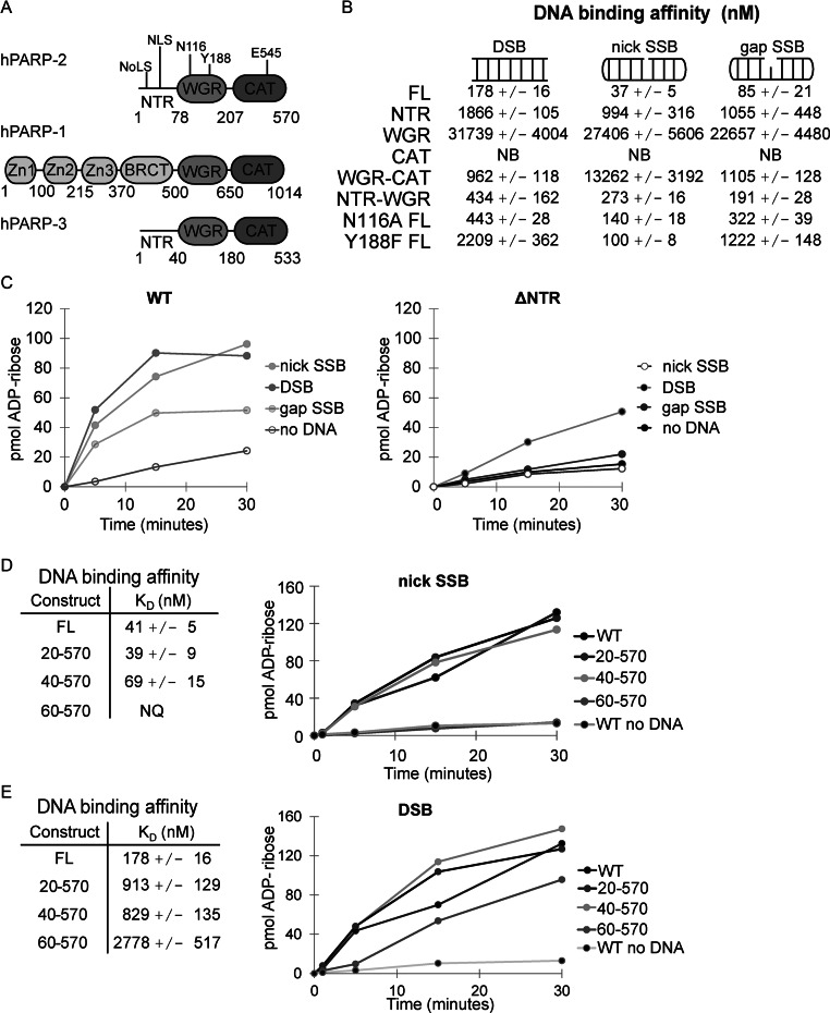
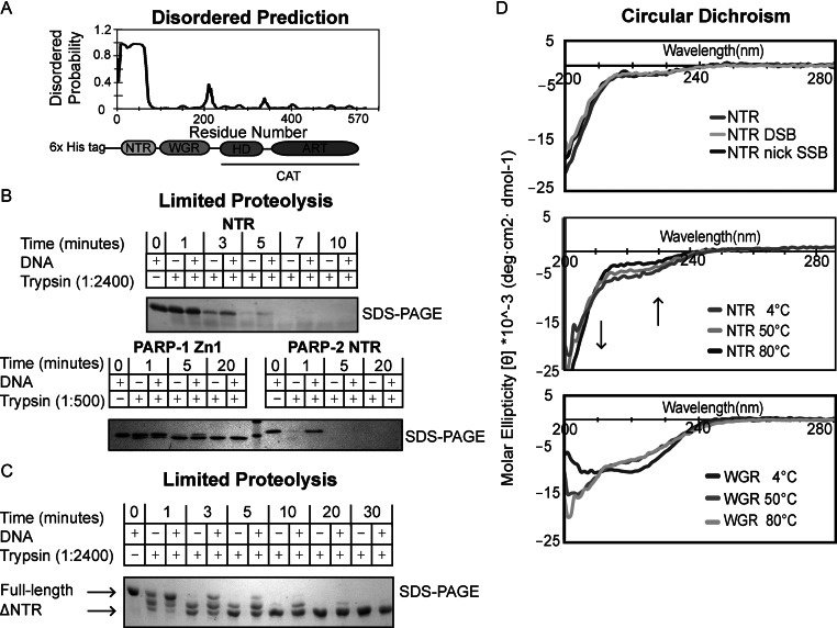
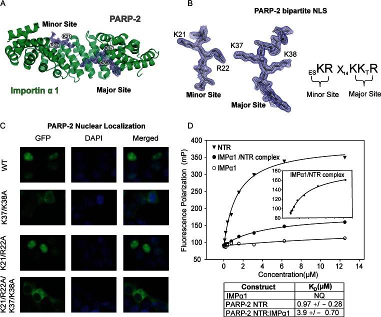
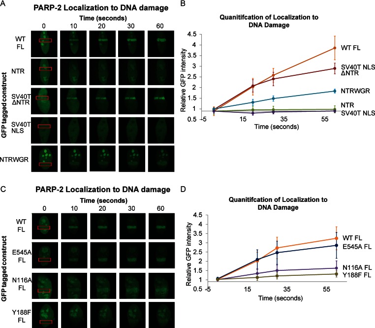
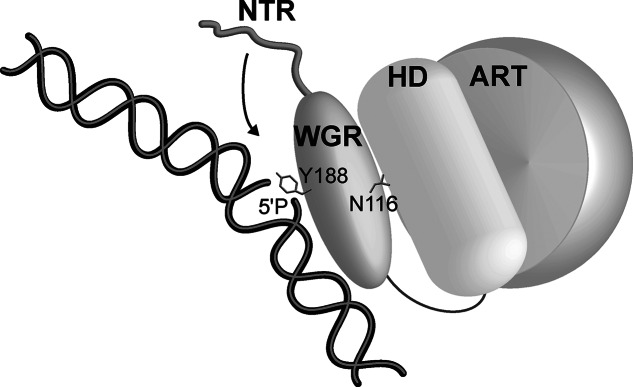
Poly(ADP-ribose) polymerase-2 (PARP-2) is one of three human PARP enzymes that are potently activated during the cellular DNA damage response (DDR). DDR-PARPs detect DNA strand breaks, leading to a dramatic increase in their catalytic production of the posttranslational modification poly(ADP-ribose) (PAR) to facilitate repair. There are limited biochemical and structural insights into the functional domains of PARP-2, which has restricted our understanding of how PARP-2 is specialized toward specific repair pathways. PARP-2 has a modular architecture composed of a C-terminal catalytic domain (CAT), a central Trp-Gly-Arg (WGR) domain and an N-terminal region (NTR). Although the NTR is generally considered the key DNA-binding domain of PARP-2, we report here that all three domains of PARP-2 collectively contribute to interaction with DNA damage. Biophysical, structural and biochemical analyses indicate that the NTR is natively disordered, and is only required for activation on specific types of DNA damage. Interestingly, the NTR is not essential for PARP-2 localization to sites of DNA damage. Rather, the WGR and CAT domains function together to recruit PARP-2 to sites of DNA breaks. Our study differentiates the functions of PARP-2 domains from those of PARP-1, the other major DDR-PARP, and highlights the specialization of the multi-domain architectures of DDR-PARPs.


Figures

Figure 1.
Figure 1.
PARP-2 NTR contributes to SSB recognition. (A) Schematic of PARP-1, PARP-2 and PARP-3 domains. Specific regions of PARP-2 are noted: NoLS (nucleolar localization signal, residues 4–7), NLS (bipartite nuclear localization signal, including K21, R22 and K36, K37), key WGR residues N116 (contact to CAT) and Y188 (contact to DNA) and a key catalytic active site residue E545 (catalytic residue). (B) The DNA-binding affinities of PARP-2 WT (1–570) and mutant constructs (NTR 1–78; WGR 71–207; CAT 216–570; WGR-CAT 71–570; NTR-WGR 1–207; N116A FL 1–570; Y188F FL 1–570) were measured by fluorescence polarization using various fluorescently labeled DNA break structures (5 nM). The KD value indicated represents the average derived from three independent experiments with associated standard deviation (SD). Proteins that showed no apparent binding are labeled NB for no binding (see also Supplemental Figures S3–S5). (C) PARP-2 DNA-dependent activity was measured using a colorimetric assay. FL PARP-2 and ΔNTR (WGR-CAT) (60 nM) were incubated with various DNA templates (480 nM). Activity data shown are representative of three independent experiments performed. (D) and (E) Left: KD of PARP-2 N-terminal truncations on a nick SSB (D) or DSB (E) template measured by fluorescence polarization. The KD indicated is the average of three independent experiments with associated SD. Right: colorimetric assay showing the activity of PARP-2 N-terminal truncation constructs (60 nM) in the presence of a nick SSB (D) or DSB (E) DNA template (480 nM). Activity data shown are representative of the three independent experiments performed.
Figure 2.
Figure 2.
The N-terminal region (NTR) of PARP-2 is natively disordered. (A) Predicted disordered regions of human PARP-2 as determined by the bioinformatics tool DISOPRED. The NTR of PARP-2 has a high probability of being disordered. (B) Time course of human PARP-2 limited proteolysis resolved on 18% SDS-PAGE. Top: SDS-PAGE analysis of 1:2400 trypsin digest of PARP-2 NTR (2 μg) in the presence and absence of DSB (2 μM) for the indicated time points. Bottom: SDS-PAGE analysis of 1:500 trypsin digest of PARP-1 Zn1 and PARP-2 NTR (2 μg) in the presence and absence of DSB (2 μM). (C) Time course of human full-length (FL) PARP-2 limited trypsin proteolysis (1:2400) resolved on 7.5% SDS-PAGE in the presence and absence of DSB (2 μM). FL PARP-2 limited proteolysis analysis on SDS-PAGE showing a truncated, proteolytic resistant PARP-2 after 5 min (D) Top: CD analysis of PARP-2 NTR data collected at 4°C using 10 μM protein in the absence or presence of activating DNA (DSB and nick SSB) (10 μM). Middle: PARP-2 NTR displays identical CD signal at 4, 50 and 80°C. Arrows indicate regions of temperature-dependent changes in CD signal expected for intrinsically disordered proteins. Bottom: temperature-dependent CD analysis of PARP-2 WGR (10 μM) showing that WGR undergoes a structural transition from 4 to 50 to 80°C. All scans were performed in triplicates and averaged to generate the curve shown. The curves are representative of three independent experiments.
Figure 3.
Figure 3.
IMPα1ΔIBB:PARP-2 bipartite NLS interaction. (A) Crystal structure of human PARP-2 NLS bound to IMPα1ΔIBB. Cartoon overview of IMPα1ΔIBB (ribbons) and PARP-2 bipartite NLS (sticks). The final σ-weighted 2FO–FC electron density map contoured at 1.5σ is overlayed. (B) Left: electron density of the PARP-2 NLS minor and major binding sites. Key PARP-2 residues are shown. Right: schematic illustration of the key PARP-2 NLS residues that mediate the interaction, where X represents any amino acid. (C) Live-cell imaging of HEK 293 cells expressing GFP-PARP-2, and mutations and truncations thereof. PARP-2 requires a bipartite NLS for maintenance of strong nuclear localization. A representative image of a population of cells is shown from three independent experiments for each GFP, DAPI and the merged DAPI/GFP image. (D) Fluorescence polarization experiment showing DNA binding of human PARP-2 NTR, IMPα1ΔIBB and the IMPα1ΔIBB/NTR complex using a fluorescently labeled 5'P nick SSB template (5 nM). Inset is a zoomed-in image showing IMPα1ΔIBB/NTR DNA-binding activity. The reported KD values are averages derived from three independent experiments with associated SD. Representative binding curves are shown.
Figure 4.
Figure 4.
PARP-2 recruitment to cellular sites of DNA damage. (A) and (C) Live-cell imaging of recruitment of GFP-PARP-2, truncation constructs (A) or FL mutants (C) to sites of laser-induced DNA damage. The region of laser irradiation is indicated with a red box. The images were captured at the various time points indicated. The images shown are representative the results obtained from at least three independent experiments. SV40T NLS is an N-terminal peptide tethered to PARP-2 truncations that lack the canonical NLS of PARP-2. (B) and (D) Quantitation of relative GFP intensity within the laser path relative to background (a non-irradiated area of the nucleus). Relative GFP signal averaged for ≥4 cells. Error bars represent the SD.
Figure 5.
Figure 5.
Model for PARP-2 recognition of DNA damage. The WGR domain forms direct contacts with DNA damage (e.g. Y188); however, WGR domain cross-talk with the HD region of the CAT domain (e.g. N116) is required to allow the domains to synergize and effectively detect DNA damage. These same contacts are necessary for DNA damage-dependent activation. The inherent flexibility of the NTR allows it to engage DNA in an adaptable manner, dependent on the nature of the damage DNA intermediate (e.g. gapped or nicked SSB).

References

    1. Gibson B.A., Kraus W.L. New insights into the molecular and cellular functions of poly(ADP-ribose) and PARPs. Nat. Rev. Mol. Cell Biol. 2012;13:411–424. - PubMed
    1. Vyas S., Chesarone-Cataldo M., Todorova T., Huang Y.-H., Chang P. A systematic analysis of the PARP protein family identifies new functions critical for cell physiology. Nat. Commun. 2013;4:2240. - PMC - PubMed
    1. Žaja R., Mikoč A., Barkauskaite E., Ahel I. Molecular insights into poly(ADP-ribose) recognition and processing. Biomolecules. 2012;3:1–17. - PMC - PubMed
    1. De Vos M., Schreiber V., Dantzer F. The diverse roles and clinical relevance of PARPs in DNA damage repair: current state of the art. Biochem. Pharmacol. 2012;84:137–146. - PubMed
    1. Kutuzov M.M., Khodyreva S.N., Amé J.C., Ilina E.S., Sukhanova M. V., Schreiber V., Lavrik O.I. Interaction of PARP-2 with DNA structures mimicking DNA repair intermediates and consequences on activity of base excision repair proteins. Biochimie. 2013;95:1208–1215. - PubMed

Publication types

MeSH terms

Substances