

Diffuse Infantile Hepatic Hemangioendothelioma With Early Central Enhancement in an Adult: A Case Report of CT and MRI Findings
- PMID: 26705232
- PMCID: PMC4697998
- DOI: 10.1097/MD.0000000000002353
Diffuse Infantile Hepatic Hemangioendothelioma With Early Central Enhancement in an Adult: A Case Report of CT and MRI Findings
Abstract
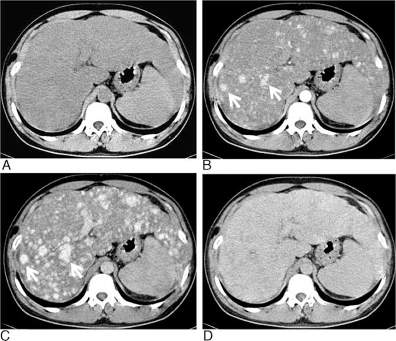
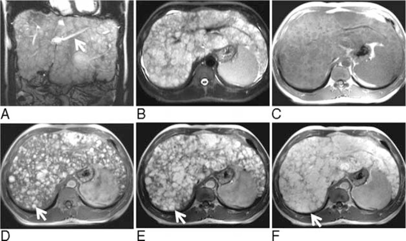
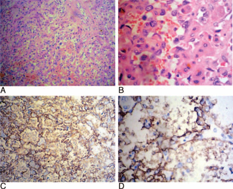
Infantile hepatic hemangioendothelioma (IHH) is the most common vascular tumor of the liver in infancy. Adult with IHH is extremely rare. We presented a diffuse IHH in an adult patient with computed tomography (CT) and magnetic resonance image (MRI) findings.A 39-year-old man was admitted to our hospital because of a 2-year history of abnormal liver function tests and a 7-day history of jaundice. Physical examination revealed enlarged liver. Unenhanced abdominal CT showed enlargement of the liver with diffuse hypodensity. Enhanced CT on the arterial phase revealed multiple centrally enhanced lesions diffusely involved the enlarged liver. The enhanced areas of the lesions became larger on the portal phase and all the lesions became homogeneous enhanced on the delayed phase. These lesions showed heterogeneously hyperintense on T2-weighted image, hypointense on T1-weighted image, and early centrally enhanced on dynamic gadolinium-enhanced MRI, with complete tumor enhancement after 180 s. The patient underwent orthotopic liver transplantation. IHH type 2 was confirmed by pathology. The patient died of tumor recurrence in the liver 4 months after transplantation.Unlike the previously described imaging appearances of IHH, this case showed diffuse nodules with early central enhancement on CT and MRI. Considering the importance of the ability to differentiate IHH from other hepatic tumors, radiologists should be aware of these imaging appearances to establish knowledge of the entire spectrum of IHH.
Conflict of interest statement
The authors have no funding and conflicts of interest to disclose.
Figures
References
-
- Keslar PJ, Buck JL, Selby DM. From the archives of the AFIP. Infantile hemangioendothelioma of the liver revisited. Radiographics 1993; 13:657–670. - PubMed
-
- Selby DM, Stocker JT, Waclawiw MA, et al. Infantile hemangioendothelioma of the liver. Hepatology 1994; 20:39–45. - PubMed
-
- Diment J, Yurim O, Pappo O. Infantile hemangioendothelioma of the liver in an adult. Arch Pathol Lab Med 2001; 125:931–932. - PubMed
-
- Dehner LP, Ishak KG. Vascular tumors of the liver in infants and children. A study of 30 cases and review of the literature. Arch Pathol 1971; 92:101–111. - PubMed
-
- Daller JA, Bueno J, Gutierrez J, et al. Hepatic hemangioendothelioma: clinical experience and management strategy. J Pediatr Surg 1999; 34:98–105. - PubMed
Publication types
MeSH terms
LinkOut - more resources
Full Text Sources
Medical
