

Different Functional and Microstructural Changes Depending on Duration of Mild Cognitive Impairment in Parkinson Disease
- PMID: 26705323
- PMCID: PMC7960312
- DOI: 10.3174/ajnr.A4626
Different Functional and Microstructural Changes Depending on Duration of Mild Cognitive Impairment in Parkinson Disease
Abstract
Background and purpose: The higher cortical burden of Lewy body and Alzheimer disease-type pathology has been reported to be associated with a faster onset of cognitive impairment of Parkinson disease. So far, there has been a few studies only about the changes of gray matter volume depending on duration of cognitive impairment in Parkinson disease. Therefore, our aim was to evaluate the different patterns of structural and functional changes in Parkinson disease with mild cognitive impairment according to the duration of parkinsonism before mild cognitive impairment.
Materials and methods: Fifty-nine patients with Parkinson disease with mild cognitive impairment were classified into 2 groups on the basis of shorter (<1 year, n = 16) and longer (≥1 year, n = 43) durations of parkinsonism before mild cognitive impairment. Fifteen drug-naïve patients with de novo Parkinson disease with intact cognition were included for comparison. Cortical thickness, Tract-Based Spatial Statistics, and seed-based resting-state functional connectivity analyses were performed. Age, sex, years of education, age at onset of parkinsonism, and levodopa-equivalent dose were included as covariates.
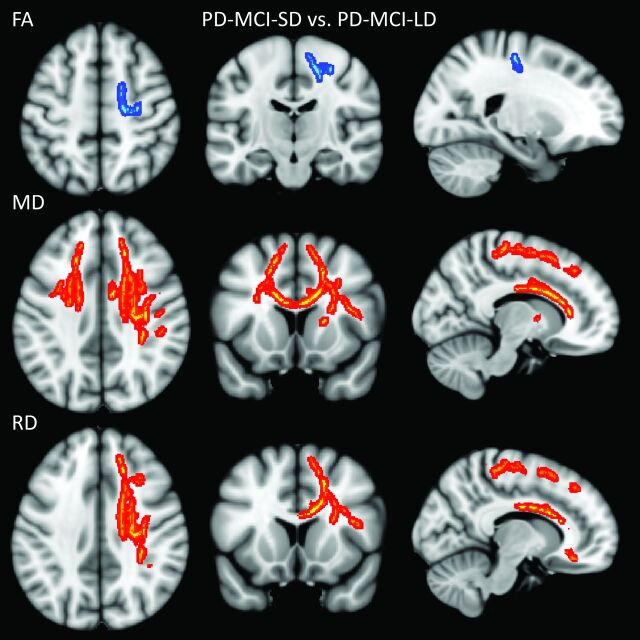
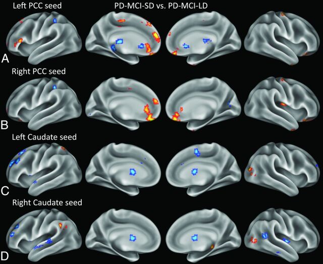
Results: The group with shorter duration of parkinsonism before mild cognitive impairment showed decreased fractional anisotropy and increased mean and radial diffusivity values in the frontal areas compared with the group with longer duration of parkinsonism before mild cognitive impairment (corrected P < .05). The group with shorter duration of parkinsonism before mild cognitive impairment showed decreased resting-state functional connectivity in the default mode network area when the left or right posterior cingulate was used as a seed, and in the dorsolateral prefrontal areas when the left or right caudate was used as a seed (corrected P < .05). The group with longer duration of parkinsonism before mild cognitive impairment showed decreased resting-state functional connectivity mainly in the medial prefrontal cortex when the left or right posterior cingulate was used as a seed, and in the parieto-occipital areas when the left or right caudate was used as a seed (corrected P < .05). No differences in cortical thickness were found in all group contrasts.
Conclusions: Resting-state functional connectivity and WM alterations might be useful imaging biomarkers for identifying changes in patients with Parkinson disease with mild cognitive impairment according to the duration of parkinsonism before mild cognitive impairment. The functional and microstructural substrates may topographically differ depending on the rate of cognitive decline in these patients.
© 2016 by American Journal of Neuroradiology.
Figures
Similar articles
-
Functional and structural changes in gray matter of parkinson's disease patients with mild cognitive impairment.Eur J Radiol. 2017 Aug;93:16-23. doi: 10.1016/j.ejrad.2017.05.018. Epub 2017 May 21. Eur J Radiol. 2017. PMID: 28668411
-
Altered functional connectivity in the default mode network is associated with cognitive impairment and brain anatomical changes in Parkinson's disease.Parkinsonism Relat Disord. 2016 Dec;33:58-64. doi: 10.1016/j.parkreldis.2016.09.012. Epub 2016 Sep 10. Parkinsonism Relat Disord. 2016. PMID: 27659747
-
Tracking Cortical Changes Throughout Cognitive Decline in Parkinson's Disease.Mov Disord. 2020 Nov;35(11):1987-1998. doi: 10.1002/mds.28228. Epub 2020 Sep 4. Mov Disord. 2020. PMID: 32886420
-
Neural Correlates of Cognitive Impairment in Parkinson's Disease: A Review of Structural MRI Findings.Int Rev Neurobiol. 2019;144:1-28. doi: 10.1016/bs.irn.2018.09.009. Epub 2018 Oct 16. Int Rev Neurobiol. 2019. PMID: 30638452 Review.
-
Mild cognitive impairment in multiple system atrophy: a brain network disorder.J Neural Transm (Vienna). 2023 Oct;130(10):1231-1240. doi: 10.1007/s00702-023-02682-x. Epub 2023 Aug 15. J Neural Transm (Vienna). 2023. PMID: 37581647 Review.
Cited by
-
Precuneus Dysfunction in Parkinson's Disease With Mild Cognitive Impairment.Front Aging Neurosci. 2019 Jan 10;10:427. doi: 10.3389/fnagi.2018.00427. eCollection 2018. Front Aging Neurosci. 2019. PMID: 30687078 Free PMC article.
-
Abnormal resting-state functional connectivity in posterior cingulate cortex of Parkinson's disease with mild cognitive impairment and dementia.CNS Neurosci Ther. 2018 Oct;24(10):897-905. doi: 10.1111/cns.12838. Epub 2018 Mar 3. CNS Neurosci Ther. 2018. PMID: 29500931 Free PMC article.
-
Delayed orthostatic hypotension in Parkinson's disease.NPJ Parkinsons Dis. 2021 Apr 14;7(1):37. doi: 10.1038/s41531-021-00181-y. NPJ Parkinsons Dis. 2021. PMID: 33854059 Free PMC article.
-
Resting-state connectivity in neurodegenerative disorders: Is there potential for an imaging biomarker?Neuroimage Clin. 2018 Mar 16;18:849-870. doi: 10.1016/j.nicl.2018.03.013. eCollection 2018. Neuroimage Clin. 2018. PMID: 29876270 Free PMC article. Review.
-
The cholinergic contribution to the resting-state functional network in non-demented Parkinson's disease.Sci Rep. 2018 May 16;8(1):7683. doi: 10.1038/s41598-018-26075-3. Sci Rep. 2018. PMID: 29769626 Free PMC article.
References
-
- Emre M, Aarsland D, Brown R, et al. . Clinical diagnostic criteria for dementia associated with Parkinson's disease. Mov Disord 2007;22:1689–707; quiz 837 - PubMed
MeSH terms
LinkOut - more resources
Full Text Sources
Medical