

Meningiomas With Rhabdoid Features Lacking Other Histologic Features of Malignancy: A Study of 44 Cases and Review of the Literature
- PMID: 26705409
- PMCID: PMC5009417
- DOI: 10.1093/jnen/nlv006
Meningiomas With Rhabdoid Features Lacking Other Histologic Features of Malignancy: A Study of 44 Cases and Review of the Literature
Abstract
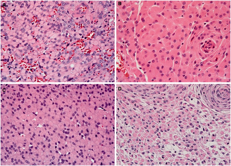
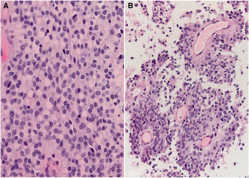
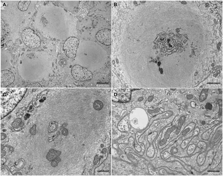
The behavior of rhabdoid meningiomas otherwise lacking malignant features remains unknown as most of the originally reported aggressive cases showed anaplastic histologic features independently of rhabdoid phenotype. We studied 44 patients with rhabdoid meningiomas lacking anaplastic features. Median age at diagnosis was 48.6 years (range 10-79). Location was supratentorial in 28 (63.6%), skull base in 15 (34.1%), and spinal in 1 (2.3%). Tumor grade was otherwise World Health Organization grade I (n = 22, 50%) or II (n = 22, 50%). Rhabdoid cells represented <20% of the tumor in 12 cases (27.3%), 20% to 50% in 18 (40.9%), and >50% in 14 (31.8%). Median clinical follow-up, available for 38 patients, was 5.0 years (range 0.17-14.2). Recurrence occurred in 9 patients (5-year recurrence-free survival, 73.7%) with a significantly higher risk in subtotally resected tumors (p = 0.043). Rhabdoid cell percentage was not associated with recurrence. Six patients died (4 of disease, 2 of unclear causes); 5-year overall survival was 86.7%, a mortality in excess of that expected in grade I-II meningiomas but much lower than originally reported. Review of 50 similar previously reported cases confirmed our findings. We suggest that rhabdoid meningiomas be graded analogously to nonrhabdoid tumors, with caution that some may still behave aggressively and close follow-up is recommended.
Keywords: Anaplastic meningioma; Meningioma; Rhabdoid meningioma; WHO grade.
© 2015 American Association of Neuropathologists, Inc. All rights reserved.
Figures
Comment in
-
Rhabdoid Meningioma: Grading and Prognostic Significance of This Uncommon Variant.J Neuropathol Exp Neurol. 2017 May 1;76(5):414-416. doi: 10.1093/jnen/nlx022. J Neuropathol Exp Neurol. 2017. PMID: 28521039 No abstract available.
References
-
- Kepes JJ, Moral LA, Wilkinson SB, et al. Rhabdoid transformation of tumor cells in meningiomas: A histologic indication of increased proliferative activity: Report of four cases. Am J Surg Pathol 1998;22:231–8 - PubMed
-
- Perry A, Scheithauer BW, Stafford SL, et al. “Rhabdoid” meningioma: An aggressive variant. Am J Surg Pathol 1998;22:1482–90 - PubMed
-
- Louis DN, Scheithauer BW, Budka H, et al. Meningiomas In: Kleihues P, Cavenee WK, eds. WHO Pathology and Genetics of Tumours of the Nervous System. Lyon: IARC Press; 2000:176–83
