Metformin Promotes Apoptosis but Suppresses Autophagy in Glucose-Deprived H4IIE Hepatocellular Carcinoma Cells
- PMID: 26706918
- PMCID: PMC4696989
- DOI: 10.4093/dmj.2015.39.6.518
Metformin Promotes Apoptosis but Suppresses Autophagy in Glucose-Deprived H4IIE Hepatocellular Carcinoma Cells
Abstract
Background: Metformin, a well-known anti-diabetic drug, has gained interest due to its association with the reduction of the prevalence of cancer in patients with type 2 diabetes and the anti-proliferative effect of metformin in several cancer cells. Here, we investigated the anti-proliferative effect of metformin with respect to apoptosis and autophagy in H4IIE hepatocellular carcinoma cells.
Methods: H4IIE rat cells were treated with metformin in glucose-free medium for 24 hours and were then subjected to experiments examining the onset of apoptosis and/or autophagy as well as the related signaling pathways.
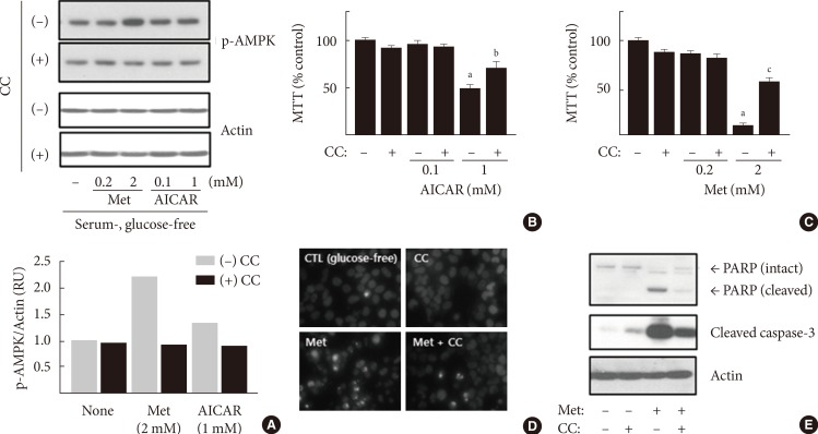
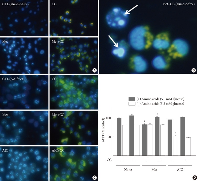
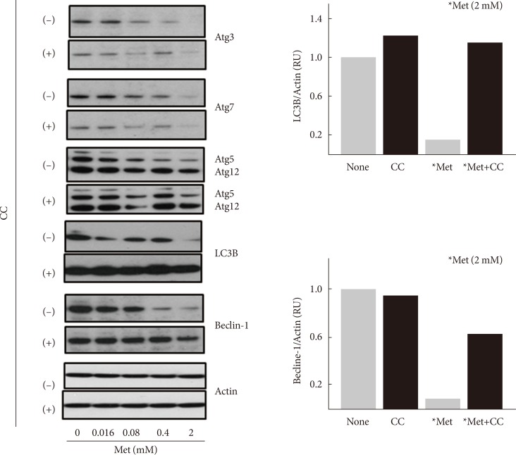
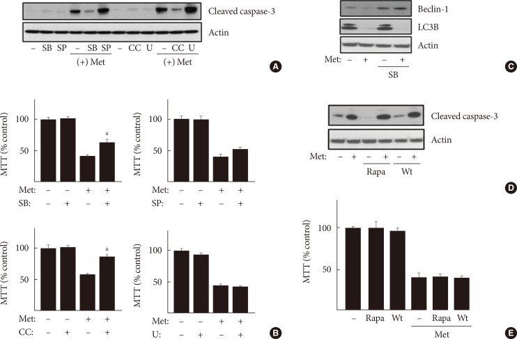
Results: When H4IIE cells were incubated in glucose-free media for 24 hours, metformin and 5-aminoimidazole-4-carboxamide ribonucleotide (AICAR) reduced the viability of cells. Inhibition of AMP-activated protein kinase (AMPK) by compound C significantly blocked cell death induced by metformin or AICAR. Pro-apoptotic events (nuclear condensation, hydrolysis of intact poly ADP ribose polymerase and caspase-3) were stimulated by metformin and then suppressed by compound C. Interestingly, the formation of acidic intracellular vesicles, a marker of autophagy, was stimulated by compound C. Although the deprivation of amino acids in culture media also induced apoptosis, neither metformin nor compound C affected cell viability. The expression levels of all of the autophagy-related proteins examined decreased with metformin, and two proteins (light chain 3 and beclin-1) were sensitive to compound C. Among the tested inhibitors against MAP kinases and phosphatidylinositol-3-kinase/mammalian target of rapamycin, SB202190 (against p38MAP kinase) significantly interrupted the effects of metformin.
Conclusion: Our data suggest that metformin induces apoptosis, but suppresses autophagy, in hepatocellular carcinoma cells via signaling pathways, including AMPK and p38 mitogen-activated protein kinase.
Keywords: Apoptosis; Autophagy; H4IIE hepatocellular carcinoma cells; Metformin.
Conflict of interest statement
Figures
Similar articles
-
Metformin Induces Lipogenesis and Apoptosis in H4IIE Hepatocellular Carcinoma Cells.Dev Reprod. 2023 Jun;27(2):77-89. doi: 10.12717/DR.2023.27.2.77. Epub 2023 Jun 30. Dev Reprod. 2023. PMID: 37529015 Free PMC article.
-
Metformin prevents an adaptive increase in GSH and induces apoptosis under the conditions of GSH deficiency in H4IIE cells.J Toxicol Environ Health A. 2007 Aug;70(15-16):1371-80. doi: 10.1080/15287390701434430. J Toxicol Environ Health A. 2007. PMID: 17654257
-
Metformin Induces Oxidative Stress-Mediated Apoptosis without the Blockade of Glycolysis in H4IIE Hepatocellular Carcinoma Cells.Biol Pharm Bull. 2019;42(12):2002-2008. doi: 10.1248/bpb.b19-00474. Biol Pharm Bull. 2019. PMID: 31787716
-
Pro-apoptotic and pro-autophagic effects of the Aurora kinase A inhibitor alisertib (MLN8237) on human osteosarcoma U-2 OS and MG-63 cells through the activation of mitochondria-mediated pathway and inhibition of p38 MAPK/PI3K/Akt/mTOR signaling pathway.Drug Des Devel Ther. 2015 Mar 12;9:1555-84. doi: 10.2147/DDDT.S74197. eCollection 2015. Drug Des Devel Ther. 2015. PMID: 25792811 Free PMC article.
-
Statins and metformin for chemoprevention of hepatocellular carcinoma.Clin Liver Dis (Hoboken). 2016 Aug 29;8(2):48-52. doi: 10.1002/cld.568. eCollection 2016 Aug. Clin Liver Dis (Hoboken). 2016. PMID: 31041062 Free PMC article. Review. No abstract available.
Cited by
-
Stable self-assembled oral metformin-bridged nanocochleates against hepatocellular carcinoma.Drug Deliv Transl Res. 2025 Jun;15(6):2064-2086. doi: 10.1007/s13346-024-01724-5. Epub 2024 Nov 13. Drug Deliv Transl Res. 2025. PMID: 39537911 Free PMC article.
-
Metformin Induces Autophagy via the AMPK-mTOR Signaling Pathway in Human Hepatocellular Carcinoma Cells.Cancer Manag Res. 2020 Jul 14;12:5803-5811. doi: 10.2147/CMAR.S257966. eCollection 2020. Cancer Manag Res. 2020. PMID: 32765083 Free PMC article.
-
Mechanistic Insights into Autophagy-Dependent Cell Death (ADCD): A Novel Avenue for Cancer Therapy.Cells. 2025 Jul 13;14(14):1072. doi: 10.3390/cells14141072. Cells. 2025. PMID: 40710325 Free PMC article. Review.
-
Exploring the protective role of metformin and dehydrozingerone in sodium fluoride-induced neurotoxicity: evidence from prenatal rat models.3 Biotech. 2025 Feb;15(2):36. doi: 10.1007/s13205-024-04175-4. Epub 2025 Jan 8. 3 Biotech. 2025. PMID: 39790448 Free PMC article.
-
Metformin in pancreatic cancer treatment: from clinical trials through basic research to biomarker quantification.J Cancer Res Clin Oncol. 2016 Oct;142(10):2159-71. doi: 10.1007/s00432-016-2178-4. Epub 2016 May 9. J Cancer Res Clin Oncol. 2016. PMID: 27160287 Free PMC article. Review.
References
-
- Pollak MN. Investigating metformin for cancer prevention and treatment: the end of the beginning. Cancer Discov. 2012;2:778–790. - PubMed
-
- Rizos CV, Elisaf MS. Metformin and cancer. Eur J Pharmacol. 2013;705:96–108. - PubMed
-
- Ashinuma H, Takiguchi Y, Kitazono S, Kitazono-Saitoh M, Kitamura A, Chiba T, Tada Y, Kurosu K, Sakaida E, Sekine I, Tanabe N, Iwama A, Yokosuka O, Tatsumi K. Antiproliferative action of metformin in human lung cancer cell lines. Oncol Rep. 2012;28:8–14. - PubMed
-
- Kato K, Gong J, Iwama H, Kitanaka A, Tani J, Miyoshi H, Nomura K, Mimura S, Kobayashi M, Aritomo Y, Kobara H, Mori H, Himoto T, Okano K, Suzuki Y, Murao K, Masaki T. The antidiabetic drug metformin inhibits gastric cancer cell proliferation in vitro and in vivo. Mol Cancer Ther. 2012;11:549–560. - PubMed
LinkOut - more resources
Full Text Sources
Other Literature Sources
Research Materials