. 2016 Sep:108:474-84.
doi: 10.1016/j.neuropharm.2015.12.005. Epub 2015 Dec 18.

Acute and chronic ethanol exposure differentially regulate CB1 receptor function at glutamatergic synapses in the rat basolateral amygdala

Affiliations

Acute and chronic ethanol exposure differentially regulate CB1 receptor function at glutamatergic synapses in the rat basolateral amygdala

Stacey L Robinson et al. Neuropharmacology. 2016 Sep.

Abstract

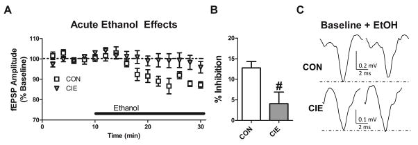
The endogenous cannabinoid (eCB) system has been suggested to play a key role in ethanol preference and intake, the acute effects of ethanol, and in the development of withdrawal symptoms following ethanol dependence. Ethanol-dependent alterations in glutamatergic signaling within the lateral/basolateral nucleus of the amygdala (BLA) are critical for the development and expression of withdrawal-induced anxiety. Notably, the eCB system significantly regulates both glutamatergic and GABAergic synaptic activity within the BLA. Chronic ethanol exposure significantly alters eCB system expression within regions critical to the expression of emotionality and anxiety-related behavior, including the BLA. Here, we investigated specific interactions between the BLA eCB system and its functional regulation of synaptic activity during acute and chronic ethanol exposure. In tissue from ethanol naïve-rats, a prolonged acute ethanol exposure caused a dose dependent inhibition of glutamatergic synaptic activity via a presynaptic mechanism that was occluded by CB1 antagonist/inverse agonists SR141716a and AM251. Importantly, this acute ethanol inhibition was attenuated following 10 day chronic intermittent ethanol vapor exposure (CIE). CIE exposure also significantly down-regulated CB1-mediated presynaptic inhibition at glutamatergic afferent terminals but spared CB1-inhibition of GABAergic synapses arising from local inhibitory-interneurons. CIE also significantly elevated BLA N-arachidonoylethanolamine (AEA or anandamide) levels and decreased CB1 receptor protein levels. Collectively, these data suggest a dynamic regulation of the BLA eCB system by acute and chronic ethanol.

Keywords: Anandamide; Basolateral amygdala; CB1; Dependence; Endogenous cannabinoid; Ethanol.


Figures

Figure 1
Figure 1
Acute ethanol dose-dependently inhibits EPSP amplitude in response to excitatory internal capsule afferent activation in the rat basolateral amygdala. A) Time course of the impact of continuous aCSF (n = 16), 20mM (n = 5), 40mM (n = 9), and 80mM (n = 6) ethanol application on fEPSP peak amplitude in drug naïve animals. B) Traces are averages of 10 minute baseline and final two minutes of treatment and illustrate significant ethanol inhibition at 40mM and 80mM doses, with no impact of continued aCSF or 20mM application. C) Dose response curve representation of percent inhibition induced by each treatment. D) 80mM ethanol increases PPR during the final five minutes of application compared to baseline, indicating a presynaptic mechanism of inhibiting excitatory transmission (n = 14). Individual points represent PPR from single cells at baseline and following acute ethanol (connected by lines). E) Traces are averages of 5 minutes baseline and the final five minutes of drug application recorded from BLA pyramidal neurons. * p < 0.05, ** p < 0.01, Dunnett’s Multiple Comparison Test; ### p < 0.001, paired t-test.
Figure 2
Figure 2
Acute ethanol inhibits excitatory internal capsule afferent terminals to rat basolateral amygdala principal neurons via the CB1 cannabinoid receptor. A) Time course of 80mM ethanol impact on fEPSP peak amplitude applied alone (n = 7) or with CB1 antagonist SR14176a (5μM, n = 6) or AM251 (1μM, n = 9). B) Traces are averages of 10 minute baseline and final two minutes of treatment. C) Bar graph representation of the percent inhibition in fEPSP peak amplitude during the final two minutes of 80mM ethanol treatment following aCSF or CB1 antagonist co-application. * p < 0.05, Dunnett’s Multiple Comparison Test.
Figure 3
Figure 3
Acute ethanol inhibition of excitatory internal capsule afferent terminals to rat basolateral amygdala is attenuated following chronic ethanol exposure. A) Time course of 80mM ethanol impact on fEPSP peak amplitude applied to drug naive control (n = 9) or chronic intermittent ethanol vapor exposed slices (n = 12). B) Bar graph representation of the percent inhibition in fEPSP peak amplitude during the final two minutes of ethanol treatment illustrating a difference in inhibition induced by 80mM ethanol between groups. C) Traces are averages of 10 minute baseline and final two minutes of treatment. # p < 0.05, t-test
Figure 4
Figure 4
Chronic ethanol exposure selectively impairs CB1 function on glutamatergic terminals in rat basolateral amygdala. A) Bar graph representation that PPR relative to baseline at glutamatergic internal capsule inputs during the final 5 minutes of WIN application is increased in CON, but not CIE cells. B) INSET: Average PPR traces for glutamatergic EPSCs at baseline (black lines) and after WIN application (gray lines) in CON and CIE cells. See text. Calibration bars are provided to denote the relative amplitude size (pA) and response duration (ms). Larger traces represent these same traces normalized for the peak amplitude of second EPSC (dashed lines) to illustrate WIN modulation of PPR. In these traces, vertical lines denote normalization effect on the relative response amplitude after scaling and are the magnitude as the inset. C) Bar graph representation that WIN increases PPR from synapses formed with GABAergic interneurons relative to baseline equally in both CON and CIE cells during the final five minutes of drug application. D) INSET: Average PPR traces for GABAergic IPSCs at baseline (black lines) and following WIN application (gray) in CON and CIE cells. Normalized traces are scaled as illustrated in B to emphasize WIN modulation of PPR. ## p < 0.01, t-test.
Figure 5
Figure 5
Chronic ethanol exposure decreases CB1 protein expression in rat basolateral amygdala. Bar graphs represent relative protein expression levels in control (CON; inset bands on left) and following chronic ethanol exposure (CIE: inset bands on right). Boxes represent approximate regions of the gel analyzed for protein expression level. See methods. A) CB1 protein expression is significantly decreased. In contrast, B) NAPE-PLD, C) FAAH, D) DAGLα, and E) MAGL expression were not significantly affected by CIE. # p < 0.05, t-test.
Figure 6
Figure 6
Chronic ethanol exposure selectively elevates tissue content of the endogenous cannabinoid AEA in rat basolateral amygdala. Bar graph representation that following chronic ethanol exposure. A) 2-AG concentrations within in the basolateral amygdala remain unchanged following chronic ethanol (CIE) relative to control (CON). In contrast, B) AEA concentration is significantly increased following chronic ethanol. # p < 0.05, t-test.
Figure 7
Figure 7
Acute ethanol inhibition of internal capsule afferent terminals to rat basolateral amygdala is not altered by FAAH inhibition. A) Time course of 80mM ethanol impact on fEPSP peak amplitude applied alone (n = 7) or following pre-incubation with the selective FAAH inhibitor URB597 (n = 7). B) Bar graph representation of the percent inhibition in fEPSP peak amplitude during the final two minutes of treatment illustrating no difference in inhibition induced by 80mM ethanol in the presence or absence of URB597. C) Traces are averages of 10 minute baseline and final two minutes of treatment.

Similar articles

Cited by

References

    1. McCool BA, Christian DT, Diaz MR, Läck AK. Glutamate plasticity in the drunken amygdala: the making of an anxious synapse. Int Rev Neurobiol. 2010;91:205–33. - PMC - PubMed
    1. Läck AK, Diaz MR, Chappell A, DuBois DW, McCool BA. Chronic ethanol and withdrawal differentially modulate pre- and postsynaptic function at glutamatergic synapses in rat basolateral amygdala. J Neurophysiol. 2007 Dec;98(6):3185–96. - PMC - PubMed
    1. Diaz MR, Christian DT, Anderson NJ, McCool BA. Chronic ethanol and withdrawal differentially modulate lateral/basolateral amygdala paracapsular and local GABAergic synapses. J Pharmacol Exp Ther. 2011 Apr;337(1):162–70. - PMC - PubMed
    1. Christian DT, Alexander NJ, Diaz MR, Robinson S, McCool BA. Chronic intermittent ethanol and withdrawal differentially modulate basolateral amygdala AMPA-type glutamate receptor function and trafficking. Neuropharmacology. 2012 Jun;62(7):2430–9. - PMC - PubMed
    1. Christian DT, Alexander NJ, Diaz MR, McCool BA. Thalamic glutamatergic afferents into the rat basolateral amygdala exhibit increased presynaptic glutamate function following withdrawal from chronic intermittent ethanol. Neuropharmacology. 2013 Feb;65:134–42. - PMC - PubMed

Publication types

MeSH terms