Review
. 2016 Feb;12(2):333-51.
doi: 10.1016/j.nano.2015.11.011. Epub 2015 Dec 17.

Biological interactions of carbon-based nanomaterials: From coronation to degradation

Affiliations
Review

Biological interactions of carbon-based nanomaterials: From coronation to degradation

Kunal Bhattacharya et al. Nanomedicine. 2016 Feb.

Abstract

Carbon-based nanomaterials including carbon nanotubes, graphene oxide, fullerenes and nanodiamonds are potential candidates for various applications in medicine such as drug delivery and imaging. However, the successful translation of nanomaterials for biomedical applications is predicated on a detailed understanding of the biological interactions of these materials. Indeed, the potential impact of the so-called bio-corona of proteins, lipids, and other biomolecules on the fate of nanomaterials in the body should not be ignored. Enzymatic degradation of carbon-based nanomaterials by immune-competent cells serves as a special case of bio-corona interactions with important implications for the medical use of such nanomaterials. In the present review, we highlight emerging biomedical applications of carbon-based nanomaterials. We also discuss recent studies on nanomaterial 'coronation' and how this impacts on biodistribution and targeting along with studies on the enzymatic degradation of carbon-based nanomaterials, and the role of surface modification of nanomaterials for these biological interactions.

From the clinical editor: Advances in technology have produced many carbon-based nanomaterials. These are increasingly being investigated for the use in diagnostics and therapeutics. Nonetheless, there remains a knowledge gap in terms of the understanding of the biological interactions of these materials. In this paper, the authors provided a comprehensive review on the recent biomedical applications and the interactions of various carbon-based nanomaterials.

Keywords: Bio-corona; Biodegradation; Carbon nanotubes; Fullerenes; Graphene oxide; Nanodiamonds.


Conflict of interest statement

Conflict of interest: The authors declare no conflicts of interest.

Figures
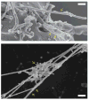
Figure 1
Figure 1
Cellular and extracellular interactions of carbon nanotubes. The upper panel shows an SEM image of isolated MWCNTs (single arrow) or a bundle of MWCNTs (two arrows) entering human mesothelial cells. Reprinted from: Shi X, von dem Bussche A, Hurt RH, Kane AB, Gao H. Cell entry of one-dimensional nanomaterials occurs by tip recognition and rotation. Nat Nanotechnol. 2011;6(11):714–9, with permission from Nature Publishing Group. The lower panel shows a cluster of short-cut SWCNTs (single arrow) entrapped in chromatin fibers (two arrows) of purified neutrophil extracellular traps [see for further details]. SEM courtesy of K. Hultenby, Karolinska Institutet.
Figure 2
Figure 2
Targeting of tumor vasculature with graphene oxide. In vivo PET/CT imaging of 64Cu-labeled GO conjugates in breast tumor-bearing mice. Left panel shows serial coronal PET images of tumor-bearing mice at different time points post-injection of 64Cu-NOTA-GO-TRC105, 64Cu-NOTA-GO, or 64Cu-NOTA-GO-TRC105 after a pre-injected blocking dose of TRC105. Tumors are indicated by arrowheads. Right panel displays representative PET/CT images of 64Cu-NOTA-GO-TRC105 in tumor-bearing mice. Reprinted from: Hong H, Yang K, Zhang Y, Engle JW, Feng L, Yang Y, Nayak TR, Goel S, Bean J, Theuer CP, Barnhart TE, Liu Z, Cai W. In vivo targeting and imaging of tumor vasculature with radiolabeled, antibody-conjugated nanographene. ACS Nano. 2012;6(3):2361–70, with permission from American Chemical Society.
Figure 3
Figure 3
Sub-organ biodistribution of carbon nanotubes. Laser desorption/ionization (LDI) mass spectrometry imaging (MSI) is an emerging label-free technique that can map chemical compounds in biological samples. From left to right: Optical image of a spleen tissue slice from mice following administration of MWCNTs; heat map showing the ion intensity distribution (m/z 72.0) of MWCNTs in a spleen tissue slice; magnified view showing the distribution of MWCNTs in the red pulp (red arrow), white pulp (white arrow) and marginal zone (purple arrow) of the spleen. Scale bars, 2 mm. Finally, representative LDI mass spectra of red and white pulp regions are depicted to the far right. Reprinted from: Chen S, Xiong C, Liu H, Wan Q, Hou J, He Q, Badu-Tawiah A, Nie Z. Mass spectrometry imaging reveals the sub-organ distribution of carbon nanomaterials. Nat Nanotechnol. 2015;10(2):176–82, with permission from Nature Publishing Group.
Figure 4
Figure 4
Enzymatic degradation of carbon nanotubes. Molecular modeling demonstrating possible SWCNT interaction sites on eosinophil peroxidase, EPO. Upper left: The two predicted interaction sites, Site 1 and Site 2 of oxidized SWCNTs modified at the edge. Upper right: Overlay of the possible interaction Site 1 of SWCNTs oxidized at the edge (colored in grey) and in the middle (colored in cyan). Lower left and right: The residues that are in close proximity (within 4 Å), stabilizing the binding sites (left) Site 1 and (right) Site 2. Positively charged residues (arginines) that are predicted to stabilize the oxidized groups on SWCNTs are colored in yellow. Reprinted from: Andón FT, Kapralov AA, Yanamala N, Feng W, Baygan A, Chambers BJ, Hultenby K, Ye F, Toprak MS, Brandner BD, Fornara A, Klein-Seetharaman J, Kotchey GP, Star A, Shvedova AA, Fadeel B, Kagan VE. Biodegradation of single-walled carbon nanotubes by eosinophil peroxidase. Small. 2013;9(16):2721–9, 2720, with permission from John Wiley and Sons.
Figure 5
Figure 5
Enzymatic degradation of graphene oxide. Upper panels show atomic force microscopy (AFM) images with section analysis of GO and horseradish peroxidase (HRP) at day 0 (left) and at day 10 (right). GO with HRP has a sheet height of 5.37 nm and 9.81 nm. Holey GO has a sheet height of 1.10 nm, and the holes were authentic at a height of 0.01 nm. Lower panels display binding poses of HRP on (from left to right) GO, holey GO, and a small sheet of GO calculated using molecular docking studies. Reprinted from: Kotchey GP, Allen BL, Vedala H, Yanamala N, Kapralov AA, Tyurina YY, Klein-Seetharaman J, Kagan VE, Star A. The enzymatic oxidation of graphene oxide. ACS Nano. 2011;5(3):2098–108, with permission from American Chemical Society.

References

    1. Fadeel B. Nanosafety: towards safer design of nanomedicines. J Intern Med. 2013;274:578–80. - PubMed
    1. Langer R, Weissleder R. Nanotechnology. JAMA. 2015;313:135–6. - PubMed
    1. Mundra RV, Wu X, Sauer J, Dordick JS, Kane RS. Nanotubes in biological applications. Curr Opin Biotechnol. 2014;28:25–32. - PubMed
    1. Chen D, Dougherty CA, Zhu K, Hong H. Theranostic applications of carbon nanomaterials in cancer: focus on imaging and cargo delivery. J Control Release. 2015;210:230–245. - PubMed
    1. Tonelli FM, Goulart VA, Gomes KN, Ladeira MS, Santos AK, Lorençon E, et al. Graphene-based nanomaterials: biological and medical applications and toxicity. Nanomedicine (Lond) 2015;10:2423–50. - PubMed

Publication types

MeSH terms