

Abnormal resting state functional connectivity in patients with chronic fatigue syndrome: an arterial spin-labeling fMRI study
- PMID: 26708036
- PMCID: PMC4801728
- DOI: 10.1016/j.mri.2015.12.008
Abnormal resting state functional connectivity in patients with chronic fatigue syndrome: an arterial spin-labeling fMRI study
Abstract
Background: Myalgic encephalomyelitis/chronic fatigue syndrome (ME/CFS) is a debilitating disorder characterized by severe fatigue and neurocognitive dysfunction. Recent work from our laboratory and others utilizing arterial spin labeling functional magnetic resonance imaging (ASL) indicated that ME/CFS patients have lower resting state regional cerebral blood flow (rCBF) in several brain areas associated with memory, cognitive, affective, and motor function. This hypoperfusion may underlie ME/CFS pathogenesis and may result in alterations of functional relationships between brain regions. The current report used ASL to compare functional connectivity of regions implicated in ME/CFS between patients and healthy controls (HC).
Methods: Participants were 17 ME/CFS patients (Mage=48.88years, SD=12) fulfilling the 1994 CDC criteria and 17 age/sex matched HC (Mage=49.82years, SD=11.32). All participants underwent T1-weighted structural MRI as well as a 6-min pseudo-continuous arterial spin labeling (pCASL) sequence, which quantifies CBF by magnetically labeling blood as it enters the brain. Imaging data were preprocessed using SPM 12 and ASL tbx, and seed-to-voxel functional connectivity analysis was conducted using the CONN toolbox. All effects noted below are significant at p<0.05 with cluster-wise FDR correction for multiple comparisons.
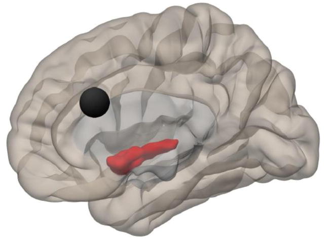
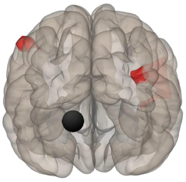
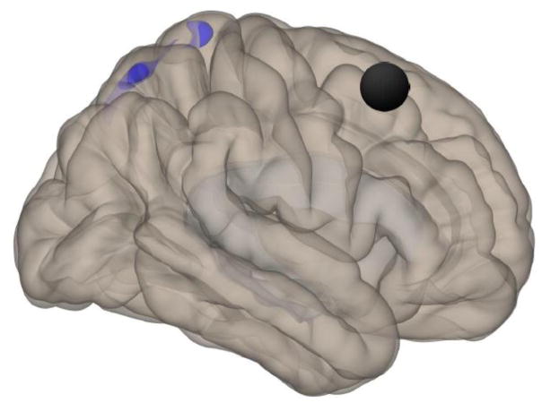
Results: ME/CFS patients demonstrated greater functional connectivity relative to HC in bilateral superior frontal gyrus, ACC, precuneus, and right angular gyrus to regions including precuneus, right postcentral gyrus, supplementary motor area, posterior cingulate gyrus, and thalamus. In contrast, HC patients had greater functional connectivity than ME/CFS in ACC, left parahippocampal gyrus, and bilateral pallidum to regions including right insula, right precentral gyrus, and hippocampus. Connectivity of the left parahippocampal gyrus correlated strongly with overall clinical fatigue of ME/CFS patients.
Conclusion: This is the first ASL based connectivity analysis of patients with ME/CFS. Our results demonstrate altered functional connectivity of several regions associated with cognitive, affective, memory, and higher cognitive function in ME/CFS patients. Connectivity to memory related brain areas (parahippocampal gyrus) was correlated with clinical fatigue ratings, providing supporting evidence that brain network abnormalities may contribute to ME/CFS pathogenesis.
Keywords: Arterial spin labeling; Chronic fatigue syndrome; Functional connectivity; MRI.
Copyright © 2015 Elsevier Inc. All rights reserved.
Figures
References
-
- Holgate ST, Komaroff AL, Mangan D, Wessely S. Chronic fatigue syndrome: understanding a complex illness. Nature Reviews Neuroscience. 2011;12(9):539–44. - PubMed
-
- Costa DC, Tannock C, Brostoff J. Brainstem perfusion is impaired in chronic fatigue syndrome. QJM. 1995;88(11):767–73. - PubMed
-
- Yoshiuchi K, Farkas J, Natelson BH. Patients with chronic fatigue syndrome have reduced absolute cortical blood flow. Clin Physiol Funct Imaging. 2006;26(2):83–6. - PubMed
-
- Staud R, Craggs JG, Lai S, Price DD, Robinson ME. Fatiguing Task is Associated with Decreased Cerebral Blood Flow in Patients with Chronic Fatigue Syndrome but not in Healthy Controls. Neuroimage Clin. 2015 in press.
Publication types
MeSH terms
Substances
Grants and funding
LinkOut - more resources
Full Text Sources
Other Literature Sources
Medical
