

doi: 10.4081/ejh.2015.2569.
Development of synovial membrane in the temporomandibular joint of the human fetus
Affiliations
- PMID: 26708184
- PMCID: PMC4698616
- DOI: 10.4081/ejh.2015.2569
Item in Clipboard
Development of synovial membrane in the temporomandibular joint of the human fetus
Eur J Histochem.
.
Abstract
The development of the synovial membrane was analyzed in serial sections of 21 temporomandibular joints of human fetuses at 9 to 13 weeks of gestation. Sections of two fetuses at 12 weeks of development were used to perform immunohistochemical expression of the markers CD68 and Hsp27 on the synovial lining. Macrophage-like type A and fibroblast-like type B cells, which express CD68 and Hsp27, respectively, were observed at the twelfth week of development. Our results suggest that the development of the synovial membrane is related to the vascularization of the joint and the formation of the articular cavities.
Conflict of interest statement
Conflict of interest: the authors declare no potential conflict of interest.
Figures
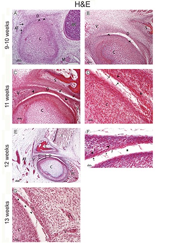
A) Human fetus (38 mm GL; week 9 of development); sagittal section stained with hematoxylin-eosin; the mandibular condyle (C) begins its chondrification; small spaces (arrowheads) show initial inferior joint cavity formation; D, articular disc; C, mandibular condyle; M, maxillary artery; AT, superficial temporal artery; MC, Meckel’s cartilage; arrows, vessel. B) Human fetus (48 mm GL; week 10 of development); frontal section stained with hematoxylin-eosin; the inferior joint cavity continues its organization between the articular disc (D) and the mandibular condyle (C); arrowheads, tracts of connective tissue; S, squamous part of the temporal bone; V, vessel. C) Human fetus (65 mm GL; week 11 of development); frontal section stained with hematoxylin-eosin; the organization of the superior joint cavity began between the squamous part of the temporal bone (S) and the articular disc (D); arrowheads, vessels in the inferior partof the primordium of the articular disc; C, mandibular condyle; V, vessels; arrows, tracts of connective tissue. D) Human fetus (65 mm GL; week 11 of development); magnification of panel A; fusiform-like cells (arrows) separate the vessels (arrowheads) of the articular disc from the inferior articular cavity; C, mandibular condyle. E) Human fetus (80 mm GL; week 12 of development); frontal section stained with hematoxylin-eosin; D, articular disc; C, mandibular condyle; PL; lateral pterygoid muscle; T, temporalis muscle; S, squamous part of the temporal bone; V, vessels; AT, superficial temporal artery. F) Human fetus (80 mm GL; week 12 of development); magnification of the squared area in panel A. Vessels (arrowheads), fusiform-like cells (arrows); inferior articular cavity (asterisk). G) Human fetus (97mm GL; week 13 of development) vessels surrounded by fusiform cells (arrowheads) form the primordium of the synovial villi; V, vessels; C, mandibular condyle.
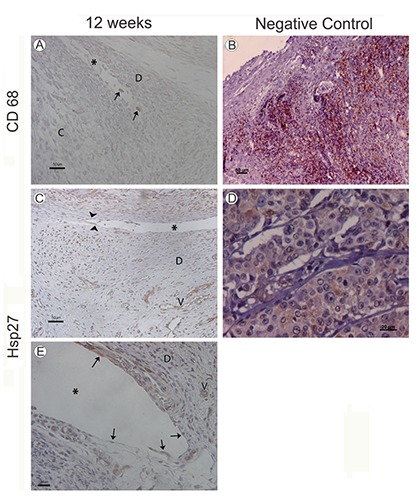
A) Human fetus (75 mm GL; week 12 of development); macrophage-like type A cells marked by CD68 antiboides; C, mandibular condyle; D, articular disc; asterisk, inferior joint cavity. B) Positive control; palatine tonsil; CD68 antibody. C) Human fetus (82 mm GL; week 12 of development); marked fibroblast-like type B cells by Hsp27 antibodies (arrowheads) in the synovial membrane and vessels (V); asterisk, superior joint cavity. D) Positive control; breast adenocarcinoma; HS27 antibody. E) Human fetus (82 mm GL; week 12 of development); marked fibroblast-like type B cells by Hsp27 antibodies (arrowheads) in the inferior articular cavity (asterisk). The walls of the vessels (V) are marked with Hsp27 antibodies; D, articular disc.
References
-
- Mérida-Velasco Jr, Rodríguez-Vázquez JF, Mérida-Velasco JA, Sánches-Montesinos I, Espín-Ferra J, et al. Development of the human temporomandibular joint. Anat Rec 1999;255:20-33. - PubMed
-
- O’Rahilly R, Müller F. Human embryology & teratology. 3rd ed. New York: Wiley-Liss; 2001.
-
- Huang GTJ, Thesleff I. Stem cells in craniofacial development and regeneration. Hoboken: Wiley Blackwell; 2013.
-
- Moraes LOC, Alvez CSR, Marques SR, Uzêda-Gonzales SQ, Vretos C, Smith RL, et al. Macroscopy and light microscopy of the discomallear passing through the petrotympanic fissure in human fetuses. Eur J Anat 2007;11:47-51.
Publication types
MeSH terms
Substances
LinkOut - more resources
Full Text Sources
Other Literature Sources
Research Materials
Miscellaneous
