

Ultrasound-guided photoacoustic imaging-directed re-endothelialization of acellular vasculature leads to improved vascular performance
- PMID: 26708553
- PMCID: PMC5988237
- DOI: 10.1016/j.actbio.2015.12.029
Ultrasound-guided photoacoustic imaging-directed re-endothelialization of acellular vasculature leads to improved vascular performance
Abstract
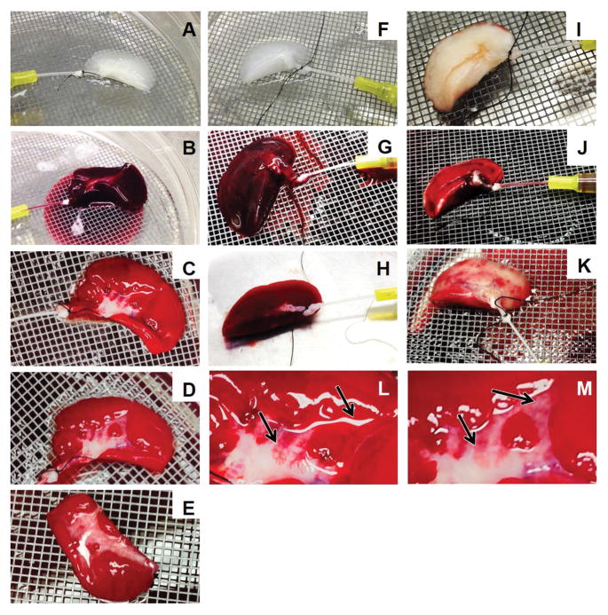
As increasing effort is dedicated to investigating the regenerative capacity of decellularized tissues, research has progressed to recellularizing these tissues prior to implantation. The delivery and support of cells seeded throughout acellular scaffolds are typically conducted through the vascular axis of the tissues. However, it is unclear how cell concentration and injection frequency can affect the distribution of cells throughout the scaffold. Furthermore, what effects re-endothelialization have on vascular patency and function are not well understood. We investigated the use of ultrasound-guided photoacoustic (US/PA) imaging as a technique to visualize the distribution of microvascular endothelial cells within an optimized acellular construct upon re-endothelialization and perfusion conditioning. We also evaluated the vascular performance of the re-endothelialized scaffold using quantitative vascular corrosion casting (qVCC) and whole-blood perfusion. We found US/PA imaging was an effective technique to visualize the distribution of cells. Cellular retention following perfusion conditioning was also detected with US/PA imaging. Finally, we demonstrated that a partial recovery of vascular performance is possible following re-endothelialization-confirmed by fewer extravasations in qVCC and improved blood clearance following whole-blood perfusion.
Statement of significance: Re-endothelialization is a method that enables decellularized tissue to become useful as a tissue engineering construct by creating a nutrient delivery and waste removal system for the entire construct. Our approach utilizes a decellularization method that retains the basement ECM of a highly vascularized tissue upon which endothelial cells can be injected to form an endothelium. The US/PA method allows for rapid visualization of cells within a construct several cm thick. This approach can be experimentally used to observe changes in cellular distribution over large intervals of time, to help optimize cell seeding parameters, and to verify cell retention within re-endothelialized constructs. This approach has temporal and depth advantages compared to section reconstruction and imaged fluorophores respectively.
Keywords: Acellular biological matrices; Angiogenesis and vasculogenesis; Bioartificial organ; Biomimetic materials; Extracellular matrix.
Copyright © 2015 Acta Materialia Inc. Published by Elsevier Ltd. All rights reserved.
Figures


Similar articles
-
Optimizing recellularization of whole decellularized heart extracellular matrix.PLoS One. 2014 Feb 27;9(2):e90406. doi: 10.1371/journal.pone.0090406. eCollection 2014. PLoS One. 2014. PMID: 24587354 Free PMC article.
-
Improving functional re-endothelialization of acellular liver scaffold using REDV cell-binding domain.Acta Biomater. 2018 Sep 15;78:151-164. doi: 10.1016/j.actbio.2018.07.046. Epub 2018 Jul 31. Acta Biomater. 2018. PMID: 30071351 Free PMC article.
-
Comparative analysis of two porcine kidney decellularization methods for maintenance of functional vascular architectures.Acta Biomater. 2018 Jul 15;75:226-234. doi: 10.1016/j.actbio.2018.06.004. Epub 2018 Jun 5. Acta Biomater. 2018. PMID: 29883813
-
Angiogenesis and Re-endothelialization in decellularized scaffolds: Recent advances and current challenges in tissue engineering.Front Bioeng Biotechnol. 2023 Feb 16;11:1103727. doi: 10.3389/fbioe.2023.1103727. eCollection 2023. Front Bioeng Biotechnol. 2023. PMID: 36873356 Free PMC article. Review.
-
Whole Cardiac Tissue Bioscaffolds.Adv Exp Med Biol. 2018;1098:85-114. doi: 10.1007/978-3-319-97421-7_5. Adv Exp Med Biol. 2018. PMID: 30238367 Review.
Cited by
-
Decellularized Human Kidney Cortex Hydrogels Enhance Kidney Microvascular Endothelial Cell Maturation and Quiescence.Tissue Eng Part A. 2016 Oct;22(19-20):1140-1150. doi: 10.1089/ten.TEA.2016.0213. Epub 2016 Aug 30. Tissue Eng Part A. 2016. PMID: 27481445 Free PMC article.
-
Dual-modal Photoacoustic and Ultrasound Imaging: from preclinical to clinical applications.Front Photon. 2024;5:1359784. doi: 10.3389/fphot.2024.1359784. Epub 2024 Feb 26. Front Photon. 2024. PMID: 39185248 Free PMC article.
-
In Vivo Tracking of Tissue Engineered Constructs.Micromachines (Basel). 2019 Jul 16;10(7):474. doi: 10.3390/mi10070474. Micromachines (Basel). 2019. PMID: 31315207 Free PMC article. Review.
-
Engineering Functional Vasculature in Decellularized Lungs Depends on Comprehensive Endothelial Cell Tropism.Front Bioeng Biotechnol. 2021 Aug 16;9:727869. doi: 10.3389/fbioe.2021.727869. eCollection 2021. Front Bioeng Biotechnol. 2021. PMID: 34485262 Free PMC article.
-
Photoacoustic Imaging in Tissue Engineering and Regenerative Medicine.Tissue Eng Part B Rev. 2020 Feb;26(1):79-102. doi: 10.1089/ten.TEB.2019.0296. Epub 2020 Jan 14. Tissue Eng Part B Rev. 2020. PMID: 31854242 Free PMC article. Review.
References
-
- Atala A, Bauer SB, Soker S, Yoo JJ, Retik AB. Tissue-engineered autologous bladders for patients needing cystoplasty. Lancet. 2006;367:1241–1246. - PubMed
-
- Brittberg M, Lindahl A, Nilsson A, Ohlsson C, Isaksson O, Peterson L. Treatment of Deep Cartilage Defects in the Knee with Autologous Chondrocyte Transplantation. N Engl J Med. 1994;331:889–895. - PubMed
-
- Kirsner RS, Falanga V, Eaglstein WH. The development of bioengineered skin. Trends Biotechnol. 1998;16:246–249. - PubMed
-
- Carmeliet P, Jain RK. Angiogenesis in cancer and other diseases. Nature. 2000;407:249–257. - PubMed
-
- Griffith CK, Miller C, Sainson RCA, Calvert JW, Jeon NL, Hughes CCW, George SC. Diffusion limits of an in vitro thick prevascularized tissue. Tissue Eng. 2005;11:257–266. - PubMed
Publication types
MeSH terms
Substances
Grants and funding
LinkOut - more resources
Full Text Sources
Other Literature Sources
Research Materials
