Review
. 2016 Apr;1862(4):814-828.
doi: 10.1016/j.bbadis.2015.12.018. Epub 2015 Dec 17.

Multiple faces of dynamin-related protein 1 and its role in Alzheimer's disease pathogenesis

Affiliations
Review

Multiple faces of dynamin-related protein 1 and its role in Alzheimer's disease pathogenesis

Ramesh Kandimalla et al. Biochim Biophys Acta. 2016 Apr.

Abstract

Mitochondria play a large role in neuronal function by constantly providing energy, particularly at synapses. Recent studies suggest that amyloid beta (Aβ) and phosphorylated tau interact with the mitochondrial fission protein, dynamin-related protein 1 (Drp1), causing excessive fragmentation of mitochondria and leading to abnormal mitochondrial dynamics and synaptic degeneration in Alzheimer's disease (AD) neurons. Recent research also revealed Aβ-induced and phosphorylated tau-induced changes in mitochondria, particularly affecting mitochondrial shape, size, distribution and axonal transport in AD neurons. These changes affect mitochondrial health and, in turn, could affect synaptic function and neuronal damage and ultimately leading to memory loss and cognitive impairment in patients with AD. This article highlights recent findings in the role of Drp1 in AD pathogenesis. This article also highlights Drp1 and its relationships to glycogen synthase kinase 3, cyclin-dependent kinase 5, p53, and microRNAs in AD pathogenesis.

Keywords: Alzheimer's disease; CDK5; Drp1; GSK3β; miRNA; mitochondria; p53.


Figures

Figure 1
Figure 1. Schematic representations of the important domains of human Drp1
Arrows on the domains are the sites of post-translational modifications. Phosphorylation, SUMOylation, S-Nirosylation were represented with the aid of arrows on Drp1 domains. The GSK3β could be interacted with GED domain of Drp1 at Ser 637 position. All amino acids were numbered with respect to human Drp1 splice variant 1 sequence (longer form).
Figure 2
Figure 2. Drp1 functions
Drp1 plays a crucial role in mitochondrial fission, fragmentation and distribution. Drp1 interacts with GSK3β and involved with the functions of synaptic plasticity across the neurons by regulating of phosphorylation of GSK3β. Drp1 also regulate the peroxisomal fragmentation, SUMOylation and ubiquitination in mammalian cells.
Figure 3
Figure 3. General overview of GSK3
GSK3 dysregulation via mitochondria with neurodegenerative diseases such as Alzheimer’s disease, Parkinson’s disease, Schizophrenia and other bipolar disorders. GSK3 also plays a regulatory role in various types of cancers, diabetes and inflammatory diseases.
Figure 4
Figure 4. Drp1 phosphorylation sites and their involvement in mitochondrial fission and fusion
(A) Phosphorylation of Drp1 at Ser637 prevents mitochondrial fragmentation; (B) conversely phosphorylation of Drp1 at Ser 616 promotes mitochondrial fragmentation. Prevention of mitochondrial fragmentation is regulated by cAMPK and promotion is carried out by CAMK1α. Drp1 is found in cytosol and cycles on/off mitochondria.
Figure 5
Figure 5. GK3β-induced Drp1 phosphorylation and mitochondrial fragmentation in Alzheimer’s disease
GSK3β-mediated phosphorylation of Drp1 at the Ser693 site in addition to Ser616 and Ser637 located within the GED domain induces elongated mitochondrial morphology which in turn inhibits mitochondrial fission/fragmentation. Blockage of GSK3β mediated Drp1 rescues Aβ induced neuronal apoptosis and provides protection from AD.
Figure 6
Figure 6. Summary of cellular and molecular changes in the pathogenesis of Alzheimer’s disease
Alzheimer’s disease is associated with the loss of synapses, oxidative stress, mitochondrial structural and functional changes, inflammatory responses, alterations in cholinergic neurotransmission, hormonal dysregulation and cell cycle abnormalities. All these changes are affected on long-term potentiation which in turn causes long term depression across the neurons, ultimately effecting synaptic transmission.
Figure 7
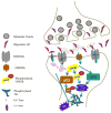
Figure 7. GS3β-Drp1-p53-CDK5 interaction in the pathogenesis of Alzheimer’s disease
Amyloid beta peptide causes hyper activation of N-methyl-D-aspartate receptors, which triggers downstream pathways such as phosphorylated tau, cyclin-dependent kinase 5, dynamin-related protein 1 and GSK-3β. These events cause endocytosis of AMPA receptors as well as NMDARs through calcium ions. These pathways may lead to abnormalities in mitochondrial dynamics and bioenergetics and impaired long-term potentiation, and synaptic dysfunction in Alzheimer’s disease neurons. The p53-dependent apoptotic pathway mobilizes the pro-apoptotic protein Bax, in turn causes mitochondrial fragmentation during apoptosis by forming a complex with Drp1 and Mfn2 at fission sites. As a transcriptional regulator, p53 may also have a direct influence on the expression and function of proteins regulating mitochondrial morphology.
Figure 8
Figure 8. Role of microRNA30 and microRNA499 in neuronal survival
Increased free radicals trigger DNA damage response and/or oxidative stress that may elicit p53-dependent apoptotic pathway. p53 mobilizes the pro-apoptotic protein Bax through the transcriptional activation of BH3 domain in proteins such as PUMA that may elevates Drp1 levels, ultimately leading to mitochondrial fragmentation. MicroRNAs, MiR30 and miR499 reduce mitochondrial fission activity by suppressing the expression of p53 and Drp1 and enhance neuronal survival.

References

    1. Yan J, Liu XH, Han MZ, Wang YM, Sun XL, Yu N, Li T, Su B, Chen ZY. Blockage of GSK3beta-mediated Drp1 phosphorylation provides neuroprotection in neuronal and mouse models of Alzheimer’s disease. Neurobiol Aging. 2015;36:211–227. - PubMed
    1. Wallace DC. Bioenergetics in human evolution and disease: implications for the origins of biological complexity and the missing genetic variation of common diseases. Philos Trans R Soc Lond B Biol Sci. 2013;368:20120267. - PMC - PubMed
    1. Reddy PH, Tripathi R, Troung Q, Tirumala K, Reddy TP, Anekonda V, Shirendeb UP, Calkins MJ, Reddy AP, Mao P, Manczak M. Abnormal mitochondrial dynamics and synaptic degeneration as early events in Alzheimer’s disease: implications to mitochondria-targeted antioxidant therapeutics. Biochim Biophys Acta. 2012;1822:639–649. - PMC - PubMed
    1. Roy M, Reddy PH, Iijima M, Sesaki H. Mitochondrial division and fusion in metabolism. Curr Opin Cell Biol. 2015;33:111–118. - PMC - PubMed
    1. Binukumar BK, Gupta N, Sunkaria A, Kandimalla R, Wani WY, Sharma DR, Bal A, Gill KD. Protective efficacy of coenzyme Q10 against DDVP-induced cognitive impairments and neurodegeneration in rats. Neurotox Res. 2012;21:345–357. - PubMed

Publication types

MeSH terms