Range of Motion Requirements for Upper-Limb Activities of Daily Living
- PMID: 26709433
- PMCID: PMC4690598
- DOI: 10.5014/ajot.2016.015487
Range of Motion Requirements for Upper-Limb Activities of Daily Living
Abstract
Objective: We quantified the range of motion (ROM) required for eight upper-extremity activities of daily living (ADLs) in healthy participants.
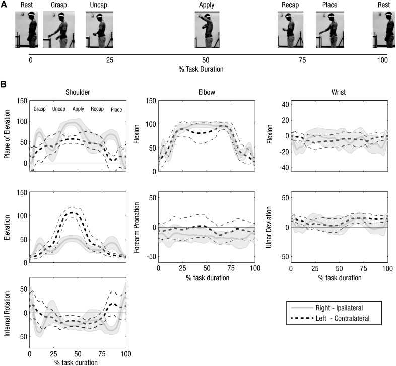
Method: Fifteen right-handed participants completed several bimanual and unilateral basic ADLs while joint kinematics were monitored using a motion capture system. Peak motions of the pelvis, trunk, shoulder, elbow, and wrist were quantified for each task.
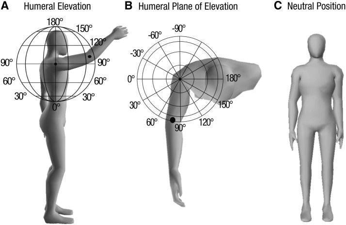
Results: To complete all activities tested, participants needed a minimum ROM of -65°/0°/105° for humeral plane angle (horizontal abduction-adduction), 0°-108° for humeral elevation, -55°/0°/79° for humeral rotation, 0°-121° for elbow flexion, -53°/0°/13° for forearm rotation, -40°/0°/38° for wrist flexion-extension, and -28°/0°/38° for wrist ulnar-radial deviation. Peak trunk ROM was 23° lean, 32° axial rotation, and 59° flexion-extension.
Conclusion: Full upper-limb kinematics were calculated for several ADLs. This methodology can be used in future studies as a basis for developing normative databases of upper-extremity motions and evaluating pathology in populations.
Copyright © 2016 by the American Occupational Therapy Association, Inc.
Figures
References
-
- Aizawa J., Masuda T., Hyodo K., Jinno T., Yagishita K., Nakamaru K., . . . Morita S. (2013). Ranges of active joint motion for the shoulder, elbow, and wrist in healthy adults. Disability and Rehabilitation, 35, 1342–1349. http://dx.doi.org/10.3109/09638288.2012.731133 - DOI - PubMed
-
- Aizawa J., Masuda T., Koyama T., Nakamaru K., Isozaki K., Okawa A., & Morita S. (2010). Three-dimensional motion of the upper extremity joints during various activities of daily living. Journal of Biomechanics, 43, 2915–2922. http://dx.doi.org/10.1016/j.jbiomech.2010.07.006 - DOI - PubMed
-
- Bonutti P. M., Windau J. E., Ables B. A., & Miller B. G. (1994). Static progressive stretch to reestablish elbow range of motion. Clinical Orthopaedics and Related Research, 303, 128–134. - PubMed
-
- Carey S. L., Jason Highsmith M., Maitland M. E., & Dubey R. V. (2008). Compensatory movements of transradial prosthesis users during common tasks. Clinical Biomechanics, 23, 1128–1135. http://dx.doi.org/10.1016/j.clinbiomech.2008.05.008 - DOI - PubMed
-
- Cutti A. G., Paolini G., Troncossi M., Cappello A., & Davalli A. (2005). Soft tissue artefact assessment in humeral axial rotation. Gait and Posture, 21, 341–349. http://dx.doi.org/10.1016/j.gaitpost.2004.04.001 - DOI - PubMed
Publication types
MeSH terms
Grants and funding
LinkOut - more resources
Full Text Sources
Other Literature Sources
Medical