. 2015 Dec 28;10(12):e0145093.
doi: 10.1371/journal.pone.0145093. eCollection 2015.

Morin, a Bioflavonoid Suppresses Monosodium Urate Crystal-Induced Inflammatory Immune Response in RAW 264.7 Macrophages through the Inhibition of Inflammatory Mediators, Intracellular ROS Levels and NF-κB Activation

Affiliations

Morin, a Bioflavonoid Suppresses Monosodium Urate Crystal-Induced Inflammatory Immune Response in RAW 264.7 Macrophages through the Inhibition of Inflammatory Mediators, Intracellular ROS Levels and NF-κB Activation

Chitra Dhanasekar et al. PLoS One. .

Abstract

Our previous studies had reported that morin, a bioflavanoid exhibited potent anti-inflammatory effect against adjuvant-induced arthritic rats. In this current study, we investigated the anti-inflammatory mechanism of morin against monosodium urate crystal (MSU)-induced inflammation in RAW 264.7 macrophage cells, an in vitro model for acute gouty arthritis. For comparison purpose, colchicine was used as a reference drug. We have observed that morin (100-300 μM) treatment significantly suppressed the levels of inflammatory cytokines (TNF-α, IL-1β, IL-6, MCP-1 and VEGF), inflammatory mediators (NO and PEG2), and lysosomal enzymes (acid phosphatase, β-galactosidase, N-acetyl glucosamindase and cathepsin D) in MSU-crystals stimulated macrophage cells. The mRNA expression of pro-inflammatory cytokines (TNF-α, IL-1β, IL-6, and MCP-1), inflammatory enzymes (iNOS and COX-2), and NF-κBp65 was found downregulated in MSU crystal stimulated macrophage cells by morin treatment, however, the mRNA expression of hypoxanthine phospho ribosyl transferse (HPRT) was found to be increased. The flow cytometry analysis revealed that morin treatment decreased intracellular reactive oxygen species levels in MSU crystal stimulated macrophage cells. The western blot analysis clearly showed that morin mainly exerts its anti-inflammatory effects by inhibiting the MSU crystal-induced COX-2 and TNF-α protein expression through the inactivation of NF-κB signaling pathway in RAW 264.7 macrophage cells similar to that of BAY 11-7082 (IκB kinase inhibitor). Our results collectively suggest that morin can be a potential therapeutic agent for inflammatory disorders like acute gouty arthritis.


Conflict of interest statement

Competing Interests: The authors have declared that no competing interests exist.

Figures

Fig 1
Fig 1. Effect of morin on the cell viability of RAW 264.7 macrophage cells.
(a) Structure of morin (b) Cells were seeded in 96-well plate and treated with varying concentration of (i) morin alone (100–500 μM) for 24 h. As well as, (ii) RAW 264.7 macrophage cells were pre-treated with varying concentration of morin (100–500 μM) followed by stimulation with MSU crystal (1 mg/ml) for 24 h. (iii) Cell phenotypes of control, MSU crystal induced and morin+MSU crystal induced RAW macrophages after 24 h were observed under phase-contrast microscope. The results are expressed as mean ±SD of the data from three experiments.
Fig 2
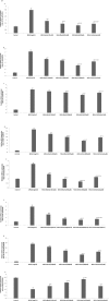
Fig 2. Effect of morin on MSU crystal-induced production of pro-inflammatory meditors in RAW 264.7 macrophage cells.
(a) The levels of (i) PGE2 and NO were measured in the culture medium. (b) The level of (i) TNF-α, (ii) IL-1β, (iii) IL-6, (iv) MCP-1, (v) VEGF and PGE2 in the culture medium was measured by its respective ELISA kits. The results are expressed as means ±SD of the data from three experiments. Comparisons are made with: a—Control versus MSU crystal-induced RAW 264.7 macrophage cells, MSU+morin-treated RAW 264.7 macrophage cells (100–300 μM) and MSU+colchicine (1 μM) treated macrophage cells; b—MSU crystal-induced RAW 264.7 macrophage cells versus MSU+morin-treated RAW 264.7 macrophage cells (100–300 μM) and MSU+colchicine (1 μM) treated RAW 264.7 macrophage cells; c, d, e—MSU+morin treated RAW 264.7 macrophage cells (100–300 μM) versus MSU+colchicine (1μM) treated RAW 264.7 macrophage cells. *P<0.05 statistically significant.
Fig 3
Fig 3. Effect of morin on MSU crystal-induced mRNA expression levels in RAW 264.7 macrophage cells.
The mRNA expression of (A) TNF-α, (B) IL-1β, (C) IL-6, (D) MCP-1, (E) iNOS, (F) COX-2, (G) NF-κBp65, (H) HPRT were measured with RT-PCR analysis. RQ values are calculated relative to the GAPDH gene. The results are expressed as mean ±SD of the data from three experiments. Comparisons are made with: a—Control versus MSU crystal-induced RAW 264.7 macrophage cells, MSU + morin treated RAW 264.7 macrophage cells (100–300 μM) and MSU+colchicine (1μM) treated RAW 264.7 macrophage cells; b—MSU crystal-induced RAW 264.7 macrophage cells versus MSU+morin treated RAW 264.7 macrophage cells (100–300 μM and MSU+colchicine (1μM) treated RAW 264.7 macrophage cells; c,d,e—MSU+morin treated RAW 264.7 macrophage cells (100–300 μM) versus MSU+colchicine (1 μM) treated RAW 264.7 macrophage cells. *P<0.05 statistically significant.
Fig 4
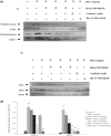
Fig 4. Effect of morin on MSU crystal induced activation of NF-κBp65, COX-2 and TNF-α in RAW 264.7 macrophage cells.
RAW 264.7 macrophage cells were treated with varying concentrations of morin (100–300 μM/ml) for 24 h and BAY 11–7028 (10 μM) for 1 h prior to the stimulation of MSU (1 mg/ml) for 24 h. Western blotting analysis was performed as described in materials and methods (i) The expression and translocation of NF-κBp65 protein were evaluated by measuring protein levels in the cytosolic and nuclear cell fractions (ii) COX-2 and TNF-α protein expression were evaluated in the whole cell lysates. β-actin and Lamin A were used as an internal loading control. The values are expressed as mean ± SEM of three independent experiments. Comparisons were made as follows: a—Control versus MSU (1mg/ml) stimulated, MSU+ morin treated groups (100–300 μM), MSU + Colchicine (1 μM); b—MSU stimulated versus MSU + morin (100–300 μM), MSU + Colchicine (1μM). *P<0.05 statistically significant, one-way ANOVA with the Bonferroni multiple comparison post-test.
Fig 5
Fig 5. Effect of morin on MSU crystal–induced intracellular ROS generation in RAW 264.7 macrophage cells.
(i) Representing the overlay image of flow cytometry. (ii) Data analysis from six experiments performed with different groups and expressed as relative fluorescence (mean ± S.D). Comparisons are made with: a—Control versus MSU crystal-induced RAW 264.7 macrophage cells, MSU+morin treated RAW 264.7 macrophage cells (100–300 μM) and MSU+colchicine (1 μM) treated RAW 264.7 macrophage cells; b—MSU crystal-induced RAW 264.7 macrophage cells versus MSU+morin treated RAW 264.7 macrophage cells (100–300 μM and MSU+colchicine (1μM) treated RAW 264.7 macrophage cells; c, d, e -MSU+morin treated RAW 264.7 macrophage cells (100–300 μM) versus MSU+colchicine (1μM) treated RAW 264.7 macrophage cells. *P<0.05 statistically significant.

References

    1. Oskouic FG, Shi Y (2013) The role of uric acid as an endogenous danger signal in immunity and inflammation. Curr Rheumatol Rep. 13(2):160–6. 10.1007/s11926-011-0162-1 . - DOI - PMC - PubMed
    1. Jin HM, Kim TJ, Choi JH, Kim MJ, Cho YN, Nam KI, et al. (2014) MicroRNA-155 as a proinflammatory regulator via SHIP-1 down-regulation in acute gouty arthritis. Arthritis Res Ther. 16(2):R88 10.1186/ar4531 . - DOI - PMC - PubMed
    1. Vanltallie TB (2010) Gout: epitome of painful arthritis. Metabolism. 1:S32–6. 10.1016/j.metabol.2010.07.009 . - DOI - PubMed
    1. Shi Y, Evans JE, Rock KL (2003) Molecular identification of a danger signal that alerts the immune system to dying cells. Nature. 425(6957):516–21. 10.1038/nature01991 . - DOI - PubMed
    1. Liu-Bryan R, Scott P, Sydlaske A, Rose DM, Terkeltaub R (2005) Innate immunity conferred by Toll-like receptors 2 and 4 and myeloid differentiation factor 88 expression is pivotal to monosodium urate monohydrate crystal-induced inflammation. Arthritis Rheum. 52(9):2936–46. 10.1002/art.21238 . - DOI - PubMed

MeSH terms

Substances