. 2015 Dec 28;10(12):e0145197.
doi: 10.1371/journal.pone.0145197. eCollection 2015.

The Macrophage Inhibitor CNI-1493 Blocks Metastasis in a Mouse Model of Ewing Sarcoma through Inhibition of Extravasation

Affiliations

The Macrophage Inhibitor CNI-1493 Blocks Metastasis in a Mouse Model of Ewing Sarcoma through Inhibition of Extravasation

Anthony J Hesketh et al. PLoS One. .

Abstract

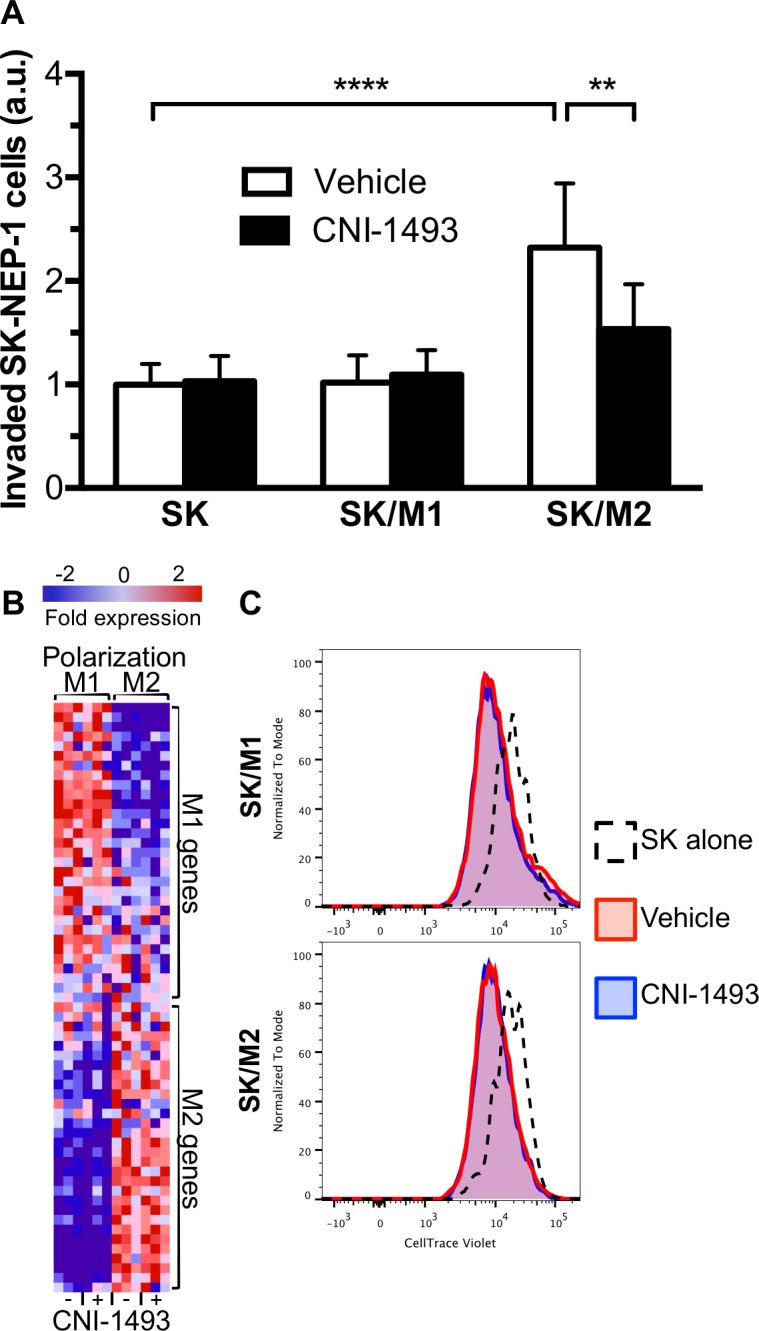
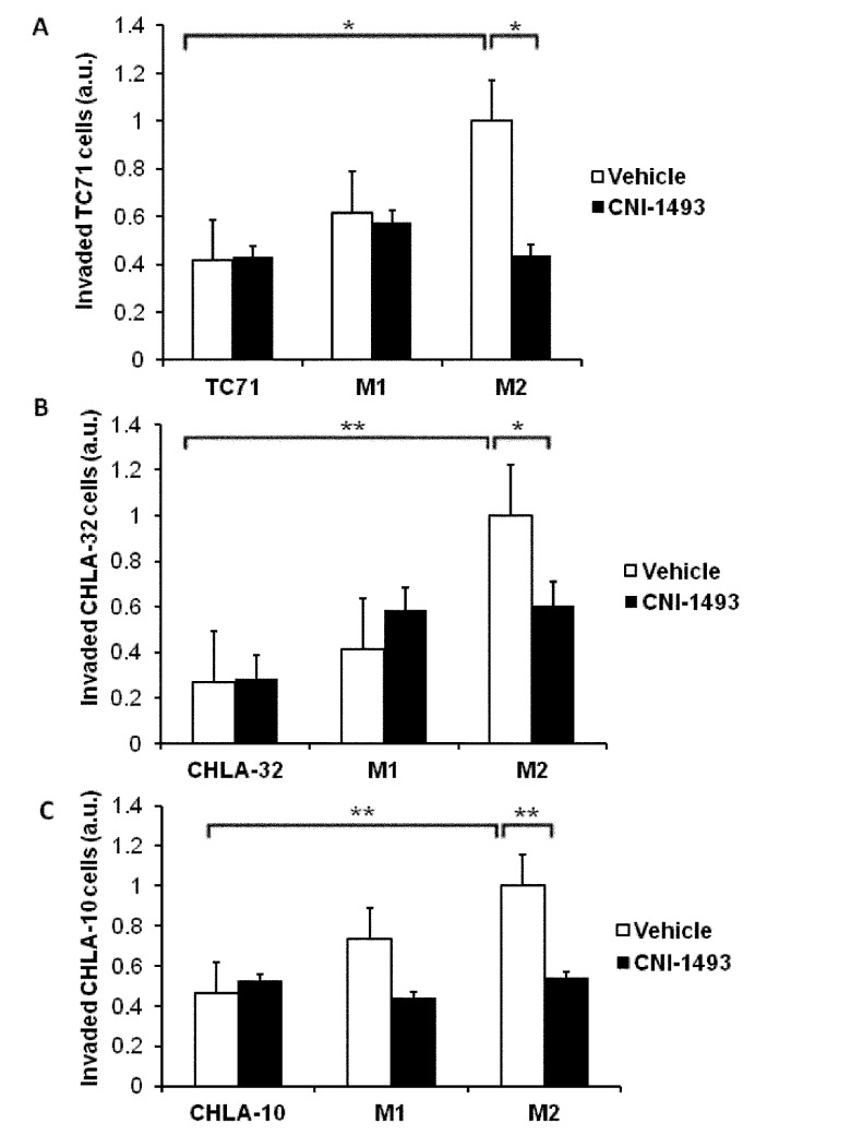
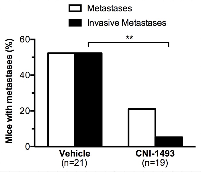
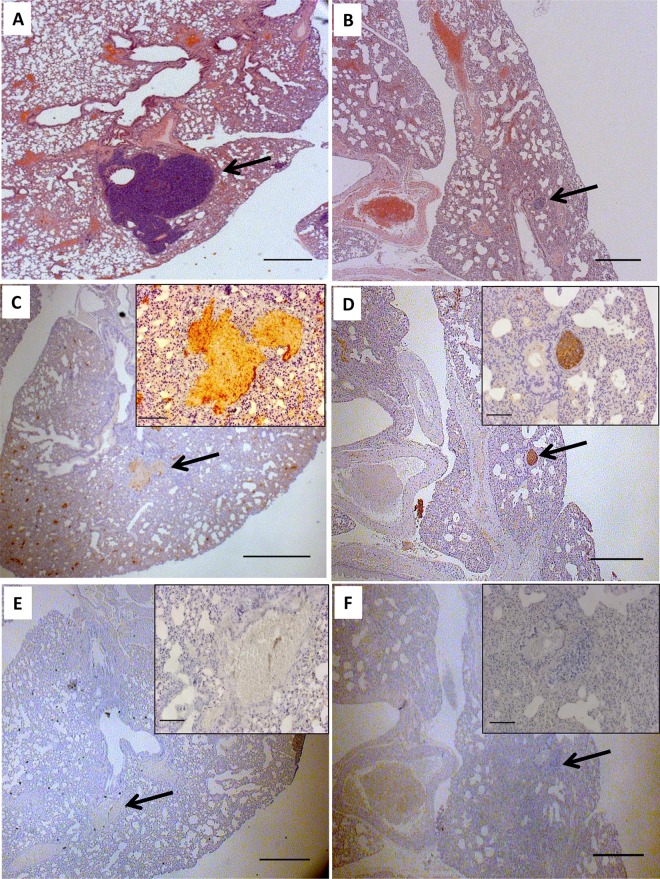
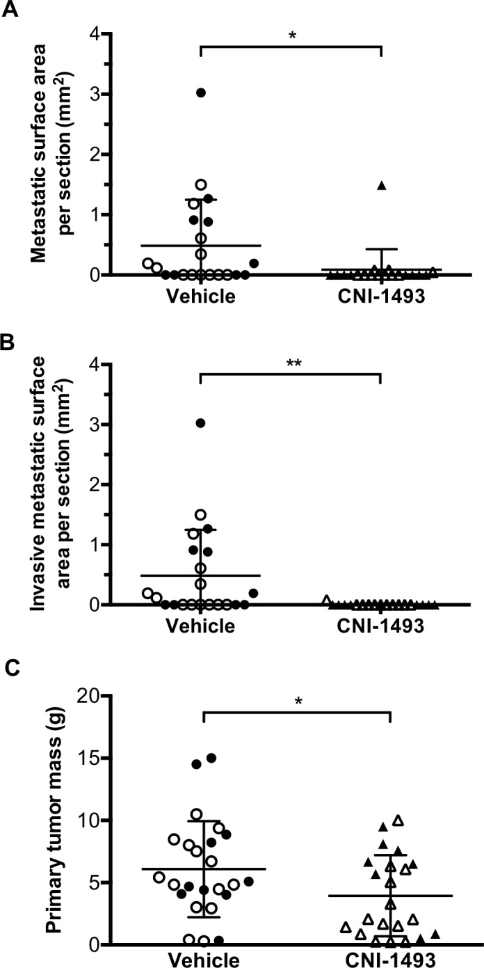
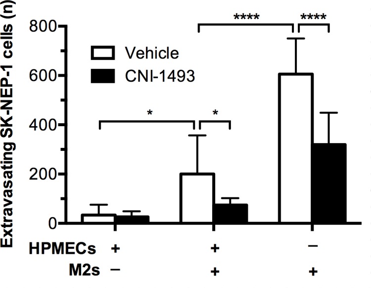
Metastatic Ewing Sarcoma carries a poor prognosis, and novel therapeutics to prevent and treat metastatic disease are greatly needed. Recent evidence demonstrates that tumor-associated macrophages in Ewing Sarcoma are associated with more advanced disease. While some macrophage phenotypes (M1) exhibit anti-tumor activity, distinct phenotypes (M2) may contribute to malignant progression and metastasis. In this study, we show that M2 macrophages promote Ewing Sarcoma invasion and extravasation, pointing to a potential target of anti-metastatic therapy. CNI-1493 is a selective inhibitor of macrophage function and has shown to be safe in clinical trials as an anti-inflammatory agent. In a xenograft mouse model of metastatic Ewing Sarcoma, CNI-1493 treatment dramatically reduces metastatic tumor burden. Furthermore, metastases in treated animals have a less invasive morphology. We show in vitro that CNI-1493 decreases M2-stimulated Ewing Sarcoma tumor cell invasion and extravasation, offering a functional mechanism through which CNI-1493 attenuates metastasis. These data indicate that CNI-1493 may be a safe and effective adjuvant agent for the prevention and treatment of metastatic Ewing Sarcoma.


Conflict of interest statement

Competing Interests: The authors have submitted a US patent application #20140323576 A1 (YA, MS, AJH, and SZS): Method for treating glioblastomas and other tumors. This application, however, does not alter the authors’ adherence to all the PLOS ONE policies on sharing data and materials.

Figures

Fig 1
Fig 1. M2-induced invasion of Ewing Sarcoma tumor cells is inhibited by CNI-1493.
(A) Fluorescently labeled SK-NEP-1 cells were cultured alone (SK) or in co-culture with M1- or M2-polarized macrophages (SK/M1 or SK/M2, respectively) in basement membrane extract within a transwell invasion culture system. Transwell systems were treated with 200 nM CNI-1493 or vehicle and the number of invading cells were counted after 72 hours. Arbitrary units represent fold change of invaded cells in vehicle-treated SK-NEP-1-only wells. **p<0.01, ****p<0.0001. Bars represent mean ± standard deviation of three experiments. (B) Gene microarray was performed on RNA extracted from M1 and M2 macrophages treated with 200 nM CNI-1493 or vehicle. Differentially expressed genes were cross-referenced with 97 previously published M1 and M2 genes as described by Martinez et al [63]. Sixty-one genes were identified and represented as a heatmap with gene expression data normalized to row. Gene names are listed in S1 Table. (C) Proliferation assay of SK-NEP-1 cells cultured alone or in co-culture with M1 or M2 macrophages (SK/M1 or SK/M2, respectively). SK-NEP-1 cells were transduced with a GFP expression vector and stained with CellTrace Violet. Cultures were treated with 200 nM CNI-1493 or vehicle and maintained in the absence of serum for 96 hours. GFP+ cells were then FACS analyzed for intensity of CellTrace Violet staining.
Fig 2
Fig 2. M2-induced invasion of Ewing Sarcoma tumor cells is inhibited by CNI-1493 in multiple cell lines.
Fluorescently labeled TC71, CHLA-32, or CHLA-10 cells were cultured alone or in co-culture with M1- or M2-polarized macrophages (labeled M1 or M2) in basement membrane extract within a transwell invasion culture system. Transwell systems were treated with 200 nM CNI-1493 or vehicle and the number of invading cells were counted after 72 hours. Arbitrary units represent fold change of invaded cells in vehicle-treated SK-NEP-1-only wells. *p<0.05, **p<0.001. Bars represent mean ± standard deviation of three experiments.
Fig 3
Fig 3. CNI-1493 reduces the incidence of invasive metastases.
Nude mice were implanted with 106 SK-NEP-1 cells in the left kidney and treated with intraperitoneal CNI-1493 (5 mg/kg/day) or vehicle for six weeks. In one replicate, drug and vehicle were delivered via subcutaneous osmotic pump placed at the time of surgery. In another replicate, drug was given via daily intraperitoneal injections. Mice were then sacrificed, lungs were sectioned, stained with hematoxylin and eosin, and examined for metastases with a pediatric pathologist in a blinded fashion. Immunohistochemistry with CD99 confirmed metastases. Metastatic foci were categorized based upon the presence of invasion into lung parenchyma. **p<0.01.
Fig 4
Fig 4. Lung sections of a metastatic Ewing Sarcoma mouse model demonstrate small, intravascular metastases in CNI-1493-treated mice.
Mice were treated as in Fig 3. Sections from vehicle-treated mice (A, C, E) demonstrated large metastases invading the lung parenchyma. The mass in Fig 4A has been serially sectioned but was still visible in 4C. Sections from CNI-1493-treated mice (B, D,F) had smaller, non-invasive metastatic tumors that were confined to the pulmonary vasculature within the endothelium. Images are representative of typical metastases, arrows indicate metastatic tumor. A, B: Hematoxylin and eosin stain, bars represent 200 μm. C, D: Immunohistochemical staining for CD99, bars represent 200 μm. E,F: Negative control for CD99 staining, bars represent 200 μm. Inset bars represent 20 μm.
Fig 5
Fig 5. CNI-1493 significantly reduced metastatic tumor burden.
Mice were treated as in Fig 3. Total cross sectional area of (A) any metastatic tumor, or (B) metastatic tumors demonstrating invasive morphology per tissue section was calculated for each mouse. (C) Primary tumors were excised at the time of animal sacrifice and weighed. Closed symbols represent the animal study replicate with intraperitoneal osmotic pump drug delivery, open symbols represent the animal study replicate with daily intraperitoneal injections. *p<0.05, **p<0.01, bars represent mean ± standard deviation.
Fig 6
Fig 6. M2-induced tumor cell extravasation across an endothelial monolayer is inhibited by CNI-1493.
GFP-expressing SK-NEP-1 cells were cultured alone or with M2 macrophages in the upper chamber of a transwell extravasation assay system, with or without the presence of an HPMEC monolayer. After 72 hours, the number of SK-NEP-1 cells invading into the lower chamber was counted. *p<0.05, ****p<0.0001. Bars represent mean ± standard deviation of four experiments.

References

    1. Balamuth NJ, Womer RB. Ewing's sarcoma. The Lancet Oncology. 2010;11(2):184–92. 10.1016/S1470-2045(09)70286-4 - DOI - PubMed
    1. Potratz J, Dirksen U, Jurgens H, Craft A. Ewing sarcoma: clinical state-of-the-art. Pediatric hematology and oncology. 2012;29(1):1–11. 10.3109/08880018.2011.622034 - DOI - PubMed
    1. Wynn TA, Chawla A, Pollard JW. Macrophage biology in development, homeostasis and disease. Nature. 2013;496(7446):445–55. 10.1038/nature12034 - DOI - PMC - PubMed
    1. Mantovani A, Sozzani S, Locati M, Allavena P, Sica A. Macrophage polarization: tumor-associated macrophages as a paradigm for polarized M2 mononuclear phagocytes. Trends in immunology. 2002;23(11):549–55. - PubMed
    1. Biswas SK, Allavena P, Mantovani A. Tumor-associated macrophages: functional diversity, clinical significance, and open questions. Seminars in immunopathology. 2013;35(5):585–600. 10.1007/s00281-013-0367-7 - DOI - PubMed

Publication types