. 2015 Dec 28;10(12):e0144991.
doi: 10.1371/journal.pone.0144991. eCollection 2015.

Identification of Thyroid Hormones and Functional Characterization of Thyroid Hormone Receptor in the Pacific Oyster Crassostrea gigas Provide Insight into Evolution of the Thyroid Hormone System

Affiliations

Identification of Thyroid Hormones and Functional Characterization of Thyroid Hormone Receptor in the Pacific Oyster Crassostrea gigas Provide Insight into Evolution of the Thyroid Hormone System

Wen Huang et al. PLoS One. .

Abstract

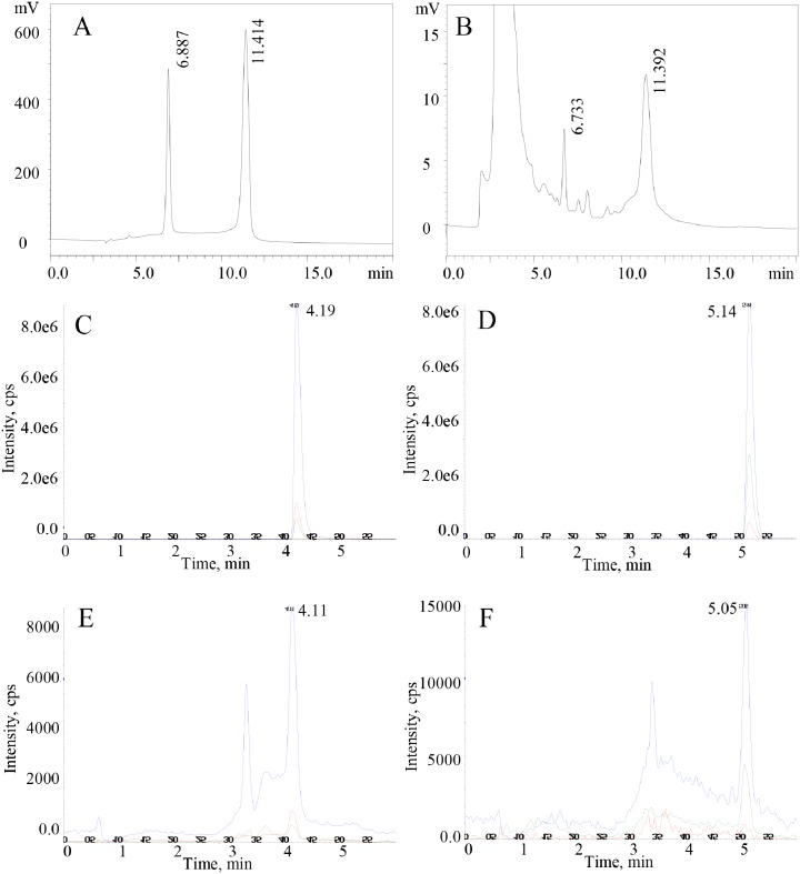
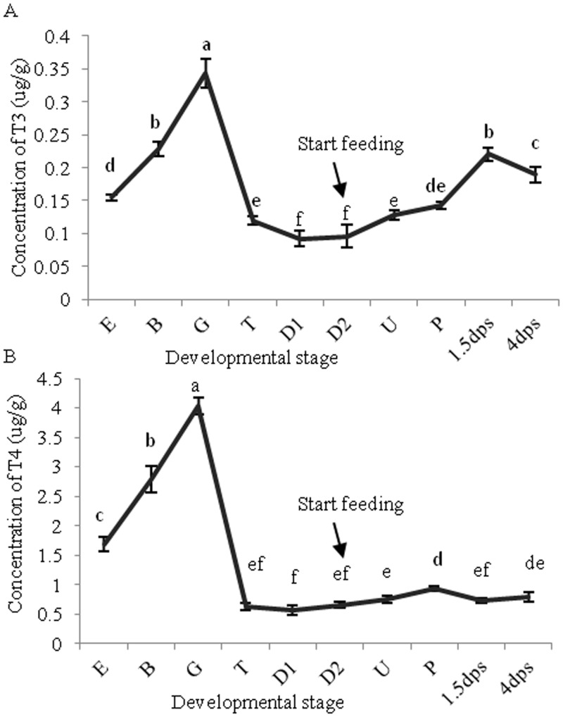
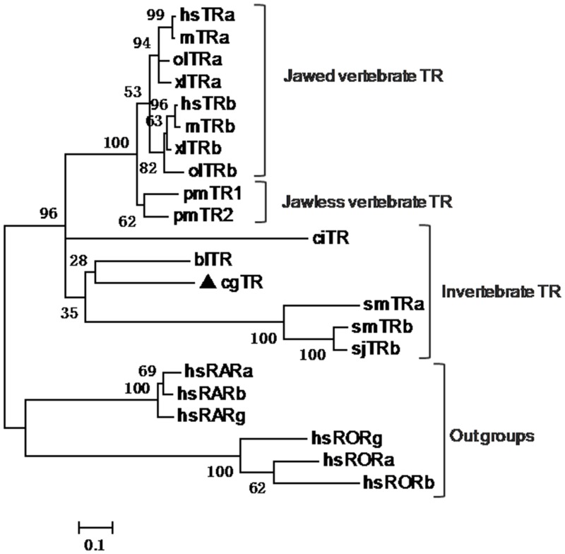
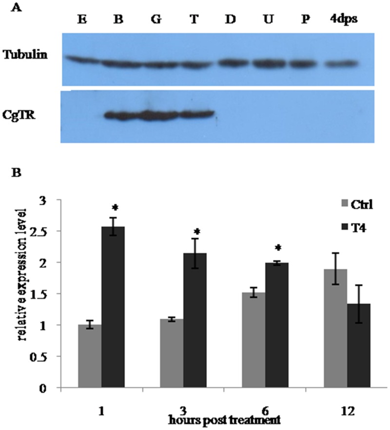
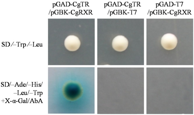
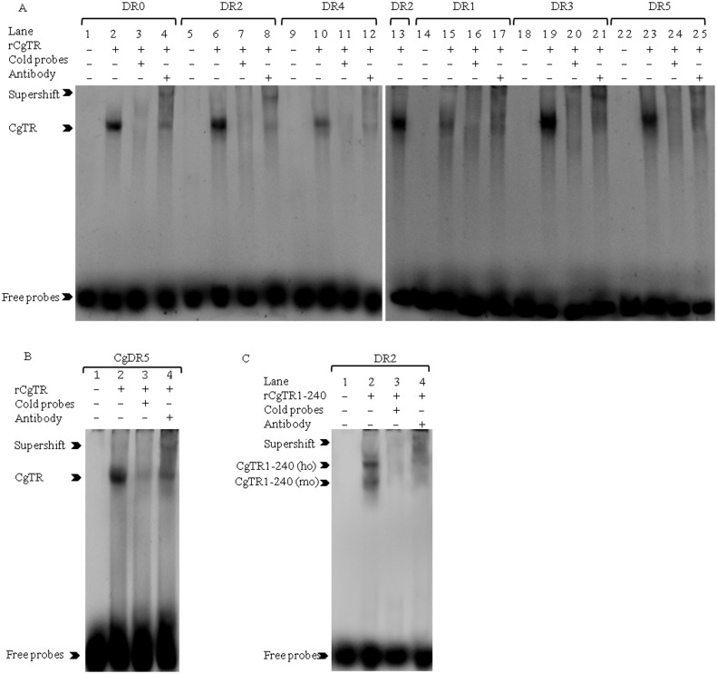
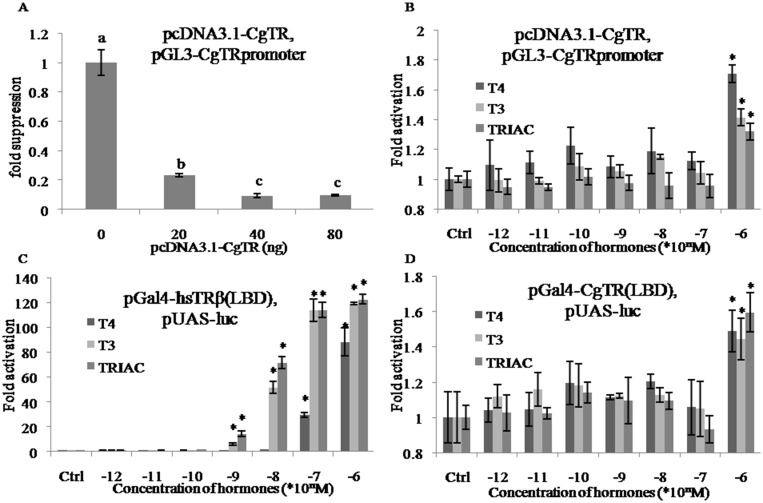
Thyroid hormones (THs) play important roles in development, metamorphosis, and metabolism in vertebrates. During the past century, TH functions were regarded as a synapomorphy of vertebrates. More recently, accumulating evidence has gradually convinced us that TH functions also occur in invertebrate chordates. To date, however, TH-related studies in non-chordate invertebrates have been limited. In this study, THs were qualitatively detected by two reliable methods (HPLC and LC/MS) in a well-studied molluscan species, the Pacific oyster Crassostrea gigas. Quantitative measurement of THs during the development of C. gigas showed high TH contents during embryogenesis and that oyster embryos may synthesize THs endogenously. As a first step in elucidating the TH signaling cascade, an ortholog of vertebrate TH receptor (TR), the most critical gene mediating TH effects, was cloned in C. gigas. The sequence of CgTR has conserved DNA-binding and ligand-binding domains that normally characterize these receptors. Experimental results demonstrated that CgTR can repress gene expression through binding to promoters of target genes and can interact with oyster retinoid X receptor. Moreover, CgTR mRNA expression was activated by T4 and the transcriptional activity of CgTR promoter was repressed by unliganded CgTR protein. An atypical thyroid hormone response element (CgDR5) was found in the promoter of CgTR, which was verified by electrophoretic mobility shift assay (EMSA). These results indicated that some of the CgTR function is conserved. However, the EMSA assay showed that DNA binding specificity of CgTR was different from that of the vertebrate TR and experiments with two dual-luciferase reporter systems indicated that l-thyroxine, 3,3',5-triiodothyronine, and triiodothyroacetic acid failed to activate the transcriptional activity of CgTR. This is the first study to functionally characterize TR in mollusks. The presence of THs and the functions of CgTR in mollusks contribute to better understanding of the evolution of the TH system.


Conflict of interest statement

Competing Interests: The authors have declared that no competing interests exist.

Figures

Fig 1
Fig 1. Qualitative measurement of T3 and T4 by HPLC (A, B) and LC/MS (C, D, E, F).
In the HPLC assay, standard T3 and T4 were separated at 6.887 min and 11.414 min (A), respectively. Extracts from the blastula contain T3 (6.733 min) and T4 (11.392 min) (B). In LC/MS, standard T3 and T4 were separated at 4.19 min (C) and 5.14 min (D), respectively. Extracts from the trochophore contain T3 (4.11 min, E) and T4 (5.05 min, F). The blue, red and green line in Fig 1C and E represent the intensity value detected by mass spectrometry of multiple reaction monitoring ions 651.6 (Q1)/605.8 (Q3), 651.6 (Q1)/508.1 (Q3), and 651.6 (Q1)/634.8 (Q3) for T3, respectively. The blue, green and red line in Fig 1C and E represent that of multiple reaction monitoring ions 777.6 (Q1)/731.6 (Q3), 777.6 (Q1)/760.8 (Q3), and 777.6 (Q1)/633.9 (Q3) for T4, respectively. Experimental conditions are described in the text.
Fig 2
Fig 2. T3 (A) and T4 (B) content (μg/g protein) in the developmental stages of C. gigas.
Larvae started feeding on Isochrysis galbana at D2 stage. Data are presented as the mean ± SD of triplicate independent experiments and were analyzed by one-way ANOVA in SPSS. TH contents at each time point that are not significantly different from one another have been marked with the same letter (P ≥ 0.05). E: egg, B: blastula, G: gastrula, T: trochophore, D: D-shape, U: umbo, P: pediveliger, dps, days post-settlement.
Fig 3
Fig 3. Phylogenetic analysis of TR.
The phylogenetic tree was derived from amino sequences of the DNA-binding domain plus ligand-binding domain of TRs. CgTR is marked with a black triangle. The human RARs and RORs were used as out groups. Protein domains were predicted by Pfam. The phylogenetic tree was constructed using the Maximum Likelihood algorithm using MEGA5.0 software. TR, thyroid hormone receptor; RAR, retinoid acid receptor; ROR, RAR-related orphan receptors; hs, Homo sapiens (hsTRa, AB307686; hsTRb, M26747; hsRARg, M24857; hsRORa, U04897; hsRORg, U16997; hsRARa, X06614; hsRARb, X07282; hsRORb, Y08639); rn, Rattus norvegicus (rnTRa, M18028; rnTRb, J03933); xl, Xenopus laevis (xlTRa, M35343; xlTRb, M35360); ol, Oryzias latipes (olTRa, AB114860; olTRb, AB114861); pm, Petromyzon marinus (pmTR1, DQ320317; pmTR2, DQ320318); bl, Branchiostoma lanceolatum (blTR, EF672345); ci, Ciona intestinalis (ciTR, NM_001032486); sm, Schistosoma mansoni (smTRa, AY395038; smTRb, AY395039); sj, Schistosoma japonicum (sjTRb, JX111998); cg, Crassostrea gigas (cgTR, KP271450).
Fig 4
Fig 4. Expression analysis of CgTR in developmental stages and in T4 treatment.
(A) Western blot analysis of CgTR protein expression in oyster developmental stages. Tubulin was used as an internal reference. (B) Temporal expression of CgTR mRNA detected by qRT-PCR during T4 treatment. RS18 gene expression was used as an internal control. Data are normalized to the control group at 1 h and displayed as the mean ± SD of triplicate independent experiments.
Fig 5
Fig 5. Yeast two-hybrid assay showing the interaction between CgTR and CgRXR.
Cotransformants containing pGAD-CgTR/pGBK-CgRXR, pGAD-T7(empty)/pGBK-CgRXR, and pGAD-CgTR/pGBK-T7(empty) grew on the SD⁄–Trp⁄–Leu medium, but only pGAD-CgTR/pGBK-CgRXR grew on the SD/–Ade/–His/–Leu/–Trp medium plus X-α-Gal/AbA
Fig 6
Fig 6. EMSA shows the DNA binding properties of CgTR in vitro.
(A) Recombinant CgTR (rCgTR) bound to probes DR0-DR5. Lanes 1, 5, 9, 14, 18, and 22 contain the reaction mixture without rCgTR as negative controls; Lanes 2, 6, 10, 15, 19, and 23 contain rCgTR; Lanes 3, 7, 11, 16, 20, and 24 contain rCgTR with a 100 fold concentration of cold probe; Lanes 4, 8, 12, 17, 21, and 25 contain rCgTR with antibody. Lane 13 contains rCgTR as an internal reference in the second plate to compare the binding intensity of DR0-DR5. As only one band was discerned, it is difficult to determine whether the bands are a homodimer or monomer. (B) EMSA demonstrated that CgTR can bind to the CgDR5. Lane 1 contains the reaction mixture without rCgTR as a negative control; Lane 2 contain rCgTR; Lane 3 contains rCgTR with a 100 fold concentration of cold probe; Lane 4 contains rCgTR with antibody. (C) Recombinant CgTR containing amino acids from 1 to 240 (rCgTR1-240) can bind to DR2 as a homodimer (ho) and monomer (mo). Lane 1 contains the reaction mixture without rCgTR1-240 as a negative control; Lane 2 contains rCgTR1-240; Lane 3 contains rCgTR1-240 with a 100 fold concentration of cold probe; Lane 4 contains rCgTR1-240 with antibody.
Fig 7
Fig 7. Transcriptional activity of CgTR could not be activated by T4, T3 and TRIAC.
(A) Transcriptional repression of the CgTR promoter by increasing amount of pcDNA3.1-CgTR (0, 20, 40, 80 ng) in absence of THs. Multiple comparisons were analyzed by one-way ANOVA in SPSS. (B) The transcriptional regulation activity of CgTR protein was hardly activated by increasing concentration of T4, T3 and TRIAC. (C) The transcriptional regulation activity of hsTRβ LBD was activated by increasing concentration of T4, T3 and TRIAC. (D) The transcriptional regulation activity of CgTR LBD was hardly activated by increasing concentration of T4, T3 and TRIAC. Data are represented as the mean ± SD of triplicate independent experiments. In (B), (C) and (D), data were pairwise compared to the corresponding control by Student’s t test and statistically significant difference are indicated by asterisks (*p < 0.05).

Similar articles

Cited by

References

    1. Yen PM. Physiological and molecular basis of thyroid hormone action. Physiol Rev. 2001;81(3):1097–142. ISI:000169570800005. - PubMed
    1. Nakajima K, Fujimoto K, Yaoita Y. Regulation of thyroid hormone sensitivity by differential expression of the thyroid hormone receptor during Xenopus metamorphosis. Genes Cells. 2012;17(8):645–59. 10.1111/j.1365-2443.2012.01614.x - DOI - PubMed
    1. Wang L, Shao YY, Ballock RT. Thyroid hormone-mediated growth and differentiation of growth plate chondrocytes involves IGF-1 modulation of beta-Catenin signaling. J Bone Miner Res. 2010;25(5):1138–46. 10.1002/Jbmr.5 ISI:000278292100024. - DOI - PMC - PubMed
    1. Tata JR. Amphibian metamorphosis as a model for the developmental actions of thyroid hormone. Mol Cell Endocrinol. 2006;246(1–2):10–20. 10.1016/j.mce.2005.11.024 ISI:000236014100002. - DOI - PubMed
    1. Morvan-Dubois G, Demeneix BA, Sachs LM. Xenopus laevis as a model for studying thyroid hormone signalling: From development to metamorphosis. Mol Cell Endocrinol. 2008;293(1–2):71–9. 10.1016/j.mce.2008.06.012 ISI:000260322800009. - DOI - PubMed

Publication types

MeSH terms