. 2016 Feb 26;291(9):4793-802.
doi: 10.1074/jbc.M115.704064. Epub 2015 Dec 28.

Insight into the Human DNA Primase Interaction with Template-Primer

Affiliations

Insight into the Human DNA Primase Interaction with Template-Primer

Andrey G Baranovskiy et al. J Biol Chem. .

Abstract

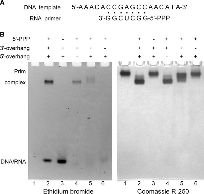
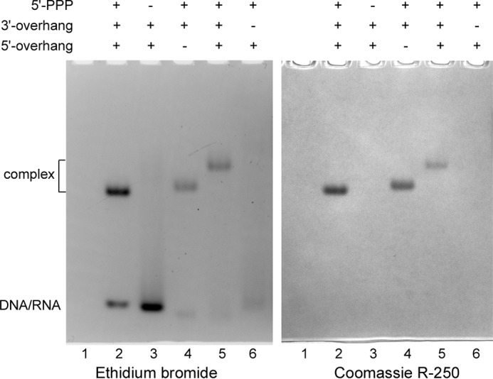
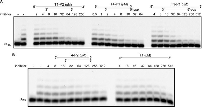
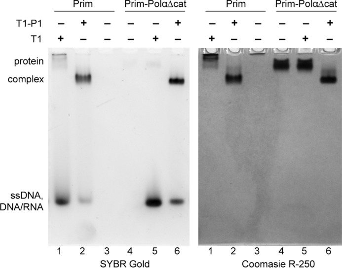
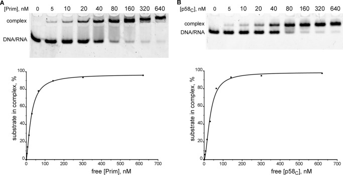
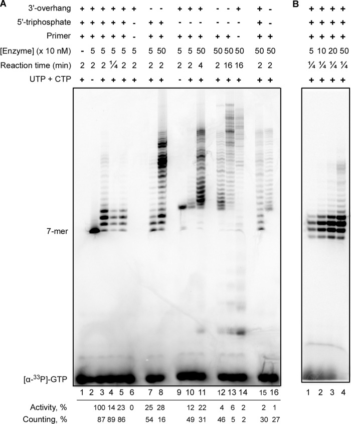
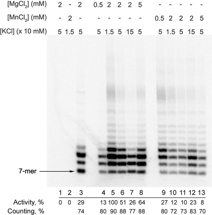
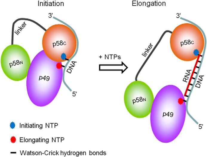
DNA replication in almost all organisms depends on the activity of DNA primase, a DNA-dependent RNA polymerase that synthesizes short RNA primers of defined size for DNA polymerases. Eukaryotic and archaeal primases are heterodimers consisting of small catalytic and large accessory subunits, both of which are necessary for the activity. The mode of interaction of primase subunits with substrates during the various steps of primer synthesis that results in the counting of primer length is not clear. Here we show that the C-terminal domain of the large subunit (p58C) plays a major role in template-primer binding and also defines the elements of the DNA template and the RNA primer that interact with p58C. The specific mode of interaction with a template-primer involving the terminal 5'-triphosphate of RNA and the 3'-overhang of DNA results in a stable complex between p58C and the DNA/RNA duplex. Our results explain how p58C participates in RNA synthesis and primer length counting and also indicate that the binding site for initiating NTP is located on p58C. These findings provide notable insight into the mechanism of primase function and are applicable for DNA primases from other species.

Keywords: 5′-triphosphate; DNA primase; DNA replication; DNA-protein interaction; RNA primer length counting; RNA synthesis; RNA/DNA duplex; gel electrophoresis; human; p58 subunit; primase activity.


Figures

FIGURE 1.
FIGURE 1.
Analysis of Prim-PolαΔcat interaction with DNA/RNA duplexes by EMSA. A, structure of the T1-P1 substrate. B, 5′-triphosphate and the 3′-overhang are critical for interaction of DNA/RNA substrate with the human primase. The total amount of Prim-PolαΔcat loaded on each of all lanes was 6 μg (30 pmol); the amount of DNA/RNA substrate in each of lanes 2–6 was 35 pmol. Lane 1, no substrate; lane 2, T1-P1; lane 3, T1-P2; lane 4, T2-P1; lane 5, T3-P1 (1-nucleotide-long 3′-overhang); lane 6, T4-P1. Samples were analyzed by electrophoresis in 5% native PAGE and stained first with ethidium bromide (left panel) and then with Coomassie R-250 (right panel). The observed difference in efficiency of DNA/RNA staining is most likely caused by effect of the template overhangs on the ethidium bromide-DNA/RNA complex in the case of short duplexes. The duplexes used in lanes 4–6 are missing the 5′- or 3′-overhang, or the overhang is shortened to one nucleotide. Reduced DNA staining in lanes 5 and 6 is also due to the smearing effect because of the protein-DNA/RNA complex dissociation during electrophoresis.
FIGURE 2.
FIGURE 2.
Analysis of p58C interaction with DNA/RNA duplexes by EMSA. The total amount of p58C loaded on each lane was 1.1 μg (50 pmol); the amount of DNA/RNA ligand in each one of lanes 2–6 was 55 pmol. Lane 1, no substrate; lane 2, T1-P1; lane 3, T1-P2; lane 4, T2-P1; lane 5, T3-P1 (1-nucleotide-long 3′-overhang); lane 6, T4-P1. Samples were analyzed by electrophoresis in 5% native PAGE and stained first with ethidium bromide (left panel) and then with Coomassie R-250 (right panel). p58C is a basic protein and moves in reverse direction when not in the complex with DNA/RNA.
FIGURE 3.
FIGURE 3.
Inhibition of rA15 primer extension by substrates with different structures. Panels A and B correspond to two representative gels. Reactions containing 40 nm Prim-PolαΔcat and 0.2 μm 5′-TYE665 fluorophore-labeled rA15 primer annealed to the dT70 template were conducted for 10 min at 30 °C. The products were analyzed by electrophoresis in 16% urea-PAGE and visualized using the Typhoon 9410 imager (emission of fluorescence at 670 nm). The left lane is the control incubation without enzyme. On the top schematics, DNA and RNA strands are shown by black and gray lines, respectively.
FIGURE 4.
FIGURE 4.
Association with PolαΔcat prevents nonspecific interaction of Prim with ssDNA. All reactions contained 5 μm protein and/or DNA. Samples were analyzed by electrophoresis in 5% native PAGE and stained first with SYBR Gold (left panel) and then with Coomassie R-250 (right panel). Prim is a basic protein and moves in reverse direction when not in the complex with ssDNA or the DNA/RNA duplex.
FIGURE 5.
FIGURE 5.
Primase and p58C bind the template-primer with a similar affinity. Primase (A) or p58C (B) of varying concentrations were incubated with T5-P1 (20 nm; T5 is labeled with Cy3 at the 5′-end). The reaction products were separated by 5% native PAGE with subsequent visualization using the Typhoon 9410 imager (emission of fluorescence at 580 nm). The graphic analyses of the obtained data were performed using the Prism 6 software (one-site specific binding with a Hill slope). Concentration of the free protein was calculated by subtracting the concentration of the protein-DNA/RNA complex from the total protein concentration.
FIGURE 6.
FIGURE 6.
Effects of the template-primer structure and p58C on primase activity. Primase activity was analyzed in reactions of extension of 6-mer RNA primers (with or without TriP) annealed with 53- or 60-mer DNA templates. The products were labeled by incorporation of [α-33P]GTP at the seventh position. Note that the negatively charged TriP moiety significantly increased the mobility of RNA primers. A, analysis of the primase activity of Prim-PolαΔcat (lanes 1–14) and PrimΔFeS-PolαΔcat (lanes 15 and 16). Lanes 1–5 and 15, T6-P3; lanes 6 and 14, T6; lanes 7 and 8, T7-P3; lanes 9–11, T6-P4; lanes 12, 13, and 16, T7-P4. Reactions corresponding to lane 5 contained 10 μm GTP. Quantification of the data is provided under each lane below the gel. The intensity of all bands was determined for estimation of the relative primase activity normalized to the protein concentration (the highest activity level was taken for 100%). The primase counting ability was calculated as the ratio of the combined intensity of the bands corresponding to the 8, 9, and 10-mer primers to the total intensity of all products, multiplied by 100%. B, effect of the enzyme-template ratio on the primase ability to terminate primer synthesis at the defined position. Reactions were run at the same conditions as for lane 4 (panel A) except the concentration of Prim-PolαΔcat was varied.
FIGURE 7.
FIGURE 7.
Effects of salt and divalent metals on primase activity and counting. Primase activity in the extension of a 6-mer RNA primer was analyzed by incubation at 35 °C of 50 nm Prim-PolαΔcat with 1 μm T6-P3 (lanes 4–13) or T8-P3 (lane 3) substrates in the presence of 100 μm each UTP and CTP and at varying salt and divalent metal concentrations. Reactions corresponding to lane 3 also contained 10 μm GTP and 100 μm ATP. The products were labeled by incorporation of [α-33P]GTP at the seventh position. Lane 1, control incubation of T6-P3 without enzyme; lane 2, same conditions as in lane 3 except the primer is absent (de novo synthesis conditions). Results of data quantification (performed as described in the legend to Fig. 6) are provided under each lane below the gel.
FIGURE 8.
FIGURE 8.
A model of interaction of the human primase with substrates at the initiation and elongation steps of primer synthesis. p58C stays bound to the initiating NTP and the 3′-overhang during the whole cycle of RNA primer synthesis, whereas p49 slides along the template toward the 5′-end.

References

    1. Loeb L. A., and Monnat R. J. Jr. (2008) DNA polymerases and human disease. Nat. Rev. Genet. 9, 594–604 - PubMed
    1. Garg P., and Burgers P. M. (2005) DNA polymerases that propagate the eukaryotic DNA replication fork. Crit. Rev. Biochem. Mol. Biol. 40, 115–128 - PubMed
    1. Pellegrini L. (2012) The Polα-primase complex. Subcell. Biochem. 62, 157–169 - PubMed
    1. Muzi-Falconi M., Giannattasio M., Foiani M., and Plevani P. (2003) The DNA polymerase α-primase complex: multiple functions and interactions. ScientificWorldJournal 3, 21–33 - PMC - PubMed
    1. Kilkenny M. L., De Piccoli G., Perera R. L., Labib K., and Pellegrini L. (2012) A conserved motif in the C-terminal tail of DNA polymerase α tethers primase to the eukaryotic replisome. J. Biol. Chem. 287, 23740–23747 - PMC - PubMed

Publication types

MeSH terms

LinkOut - more resources