. 2016 Feb 26;291(9):4589-602.
doi: 10.1074/jbc.M115.674846. Epub 2015 Dec 28.

p120RasGAP Protein Mediates Netrin-1 Protein-induced Cortical Axon Outgrowth and Guidance

Affiliations

p120RasGAP Protein Mediates Netrin-1 Protein-induced Cortical Axon Outgrowth and Guidance

Judith Antoine-Bertrand et al. J Biol Chem. .

Abstract

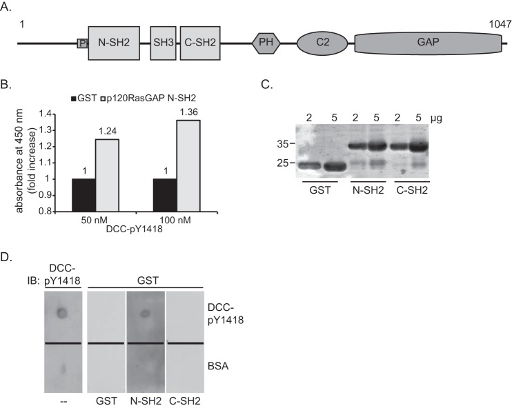
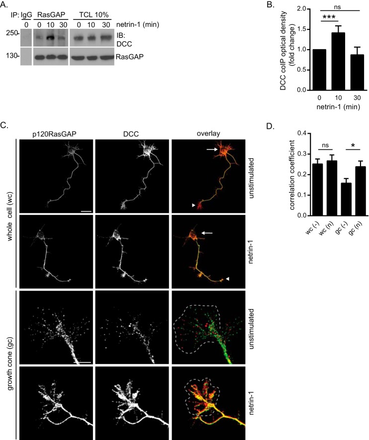
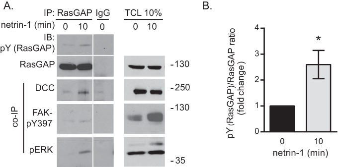
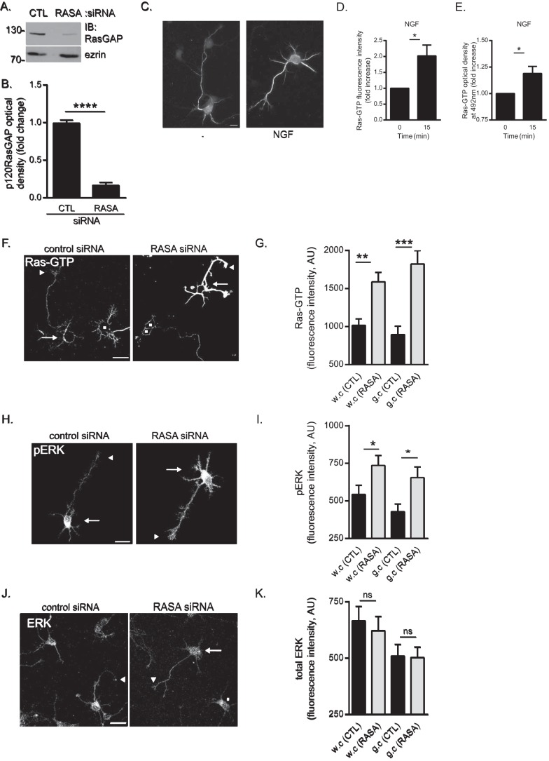
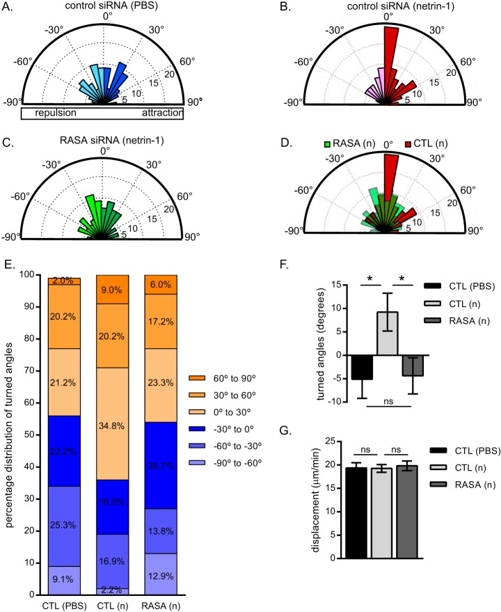
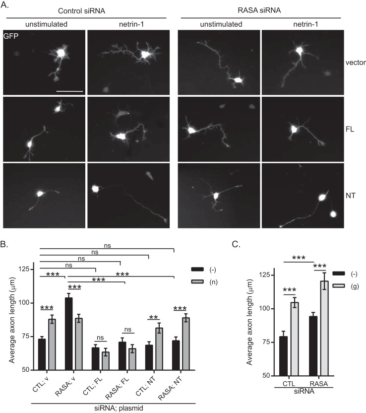
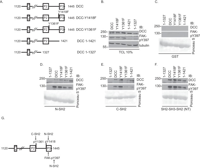
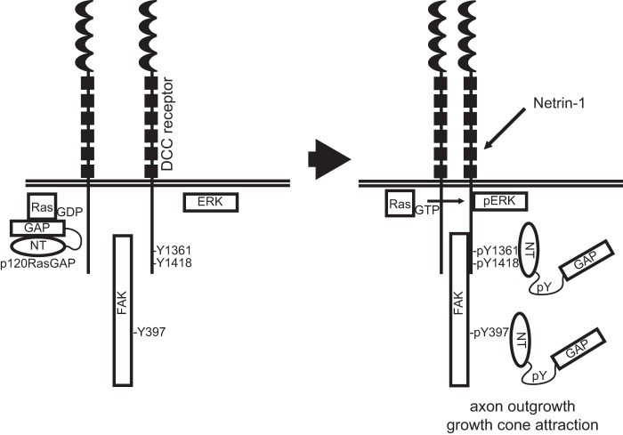
The receptor deleted in colorectal cancer (DCC) mediates the attraction of growing axons to netrin-1 during brain development. In response to netrin-1 stimulation, DCC becomes a signaling platform to recruit proteins that promote axon outgrowth and guidance. The Ras GTPase-activating protein (GAP) p120RasGAP inhibits Ras activity and mediates neurite retraction and growth cone collapse in response to repulsive guidance cues. Here we show an interaction between p120RasGAP and DCC that positively regulates netrin-1-mediated axon outgrowth and guidance in embryonic cortical neurons. In response to netrin-1, p120RasGAP is recruited to DCC in growth cones and forms a multiprotein complex with focal adhesion kinase and ERK. We found that Ras/ERK activities are elevated aberrantly in p120RasGAP-deficient neurons. Moreover, the expression of p120RasGAP Src homology 2 (SH2)-SH3-SH2 domains, which interact with the C-terminal tail of DCC, is sufficient to restore netrin-1-dependent axon outgrowth in p120RasGAP-deficient neurons. We provide a novel mechanism that exploits the scaffolding properties of the N terminus of p120RasGAP to tightly regulate netrin-1/DCC-dependent axon outgrowth and guidance.

Keywords: DCC; Ras protein; netrin-1; neurite outgrowth; neurobiology; neurodevelopment; signal transduction.


Figures

FIGURE 1.
FIGURE 1.
The N-terminal SH2 domain of p120RasGAP interacts in vitro with DCC via phosphorylated Tyr-1418. A, p120RasGAP contains a proline-rich region (P) and N-SH2 and C-SH2, SH3, pleckstrin homology (PH), calcium-dependent phospholipid-binding (C2), and GAP domains. B, DCC- Tyr(P)-1418 was used as bait to screen a SH2 domain array by ELISA. Binding of p120RasGAP N-SH2 with 50 and 100 nm of DCC- Tyr(P)-1418 peptide is represented as the -fold increase in absorbance relative to the absorbance obtained with a GST control. C, 2 and 5 μg of purified GST, GST-p120RasGAP N-SH2, and C-SH2 were resolved by SDS-PAGE, and the proteins were stained with Coomassie Blue. D, the DCC- Tyr(P)-1418 peptide was spotted onto nitrocellulose membranes with BSA as a control, and each membrane was incubated with either purified GST, GST-p120RasGAP N-SH2, or C-SH2 (100 ng/ml), followed by immunoblotting (IB) with anti-GST antibodies. One membrane was immunoblotted with phospho-specific anti-DCC-pY1418 (DCC-pY1418) antibodies.
FIGURE 2.
FIGURE 2.
p120RasGAP interacts with DCC in netrin-1-induced embryonic cortical neurons. E18 rat cortical neurons were stimulated with netrin-1 (500 ng/ml) for the indicated times after being cultured for 2 DIV. A, p120RasGAP was immunoprecipitated (IP) from cell lysates with anti-p120RasGAP antibodies or mouse IgG as a control. Immunoprecipitated proteins and total cell lysates (TCL) were resolved by SDS-PAGE and immunoblotted (IB) with antibodies against the indicated proteins. B, quantitative densitometry (mean ± S.E.) of DCC co-immunoprecipitated with p120RasGAP is represented as the -fold change relative to 0 min of netrin-1 stimulation for at least three independent experiments (unpaired Student's t test; ***, p < 0.005; ns, not significant). C, E18 embryonic rat cortical neurons (2 DIV) were incubated with 500 ng/ml netrin-1 or left unstimulated for 10 min, immunostained with antibodies against DCC and p120RasGAP, and imaged by confocal microscopy. Arrows indicate cell bodies, and arrowheads indicate growth cones. The gray dashed outlines represent growth cones. Scale bars = 50 μm (whole cells) and 20 μm (growth cones). D, the correlation between DCC and p120RasGAP fluorescence intensities in C was measured with Metamorph software using Pearson's correlation coefficient (mean ± S.E.) in whole cells (wc) and growth cones (gc) in three independent experiments (number of neurons = 65, 51, 54, and 40 from left to right; unpaired Student's t test; ns, not significant; *, p = 0.028). n, netrin-1; −, unstimulated.
FIGURE 3.
FIGURE 3.
p120RasGAP associates with a DCC multiprotein complex in netrin-1-stimulated cortical neurons. A, cortical neurons were stimulated with netrin-1 for 10 min. p120RasGAP was immunoprecipitated from cell lysates with anti-p120RasGAP antibodies or mouse IgGs as a control. Immunoprecipitated proteins and total cell lysates (TCL) were resolved by SDS-PAGE and immunoblotted (IB) with antibodies against the indicated proteins (pY (RasGAP), anti-phosphotyrosine antibodies). Data were taken from the same film exposure. The white line between the RasGAP immunoprecipitation (IP) and IgG lanes indicates that irrelevant lanes were removed digitally from the original image. B, quantitative densitometry (mean ± S.E.) of Tyr(P) (the RasGAP)/RasGAP ratio is represented as the -fold change relative to 0 min of netrin-1 stimulation for at least three independent experiments; unpaired Student's t test; *, p < 0.05).
FIGURE 4.
FIGURE 4.
p120RasGAP is required to maintain basal Ras and ERK activities in cortical neurons. Control (CTL) or p120RasGAP (RASA) siRNA was electroporated in neurons at 0 DIV with pGFP as a transfection reporter plasmid. A, total cell lysates were resolved by SDS-PAGE and immunoblotted (IB) with antibodies against p120RasGAP and ezrin as a loading control. B, quantitative densitometry of A represented as the -fold change (mean ± S.E.) relative to control siRNA measured in 10 independent experiments (unpaired Student's t test; ****, p < 0.0001). C, cortical neurons were stimulated with NGF (100 ng/ml) or left unstimulated (−) for 15 min. Neurons were immunostained with anti-Ras-GTP antibodies. Scale bar = 10 μm. D, Ras-GTP fluorescence intensity and the -fold increase (mean ± S.E.) relative to unstimulated control neurons was measured in at least three independent experiments (n > 50 neurons/condition; unpaired Student's t test; *, p < 0.05). E, the levels of Ras-GTP in each cell lysate were evaluated by G-LISA assay by measuring the absorbance at 492 nm, which is represented as the -fold change (mean ± S.E.) relative to the unstimulated lysate (0 min) in at least three independent experiments (unpaired Student's t test; *, p < 0.05). F, neurons were immunostained with anti-Ras-GTP antibodies. Arrows indicate cell bodies, arrowheads indicate growth cones of GFP-expressing neurons, and squares represent untransfected neurons. Scale bar = 50 μm. G, the Ras-GTP fluorescence intensity (arbitrary units (AU), mean ± S.E.) of GFP-expressing neurons in F was measured in whole cells (w.c) and growth cones (g.c) in three independent experiments (n = 31, 32, 31, and 32 neurons; two-way ANOVA, Fisher's least significant difference post test; **, p < 0.0004; ***, p < 0.0001). H, neurons were immunostained with antibodies against pERK. Arrows indicate cell bodies, and arrowheads indicate growth cones. Scale bar = 50 μm. I, pERK fluorescence intensity (mean ± S.E.) of GFP-expressing neurons in H was measured in whole cells and growth cones in three independent experiments (n = 35, 36, 30, and 35 neurons; two-way ANOVA, Fisher's least significant difference post test; *, p < 0.05). J, neurons were immunostained with antibodies against ERK. Arrows and arrowheads indicate cell bodies and growth cones of GFP-expressing neurons, respectively, and squares indicate untransfected neurons. Scale bar = 50 μm. K, the total ERK fluorescence intensity (mean ± S.E.) of GFP-expressing neurons in J was measured in whole cells and growth cones (n = 41, 38, 41, and 38 neurons) in three independent experiments (two-way ANOVA, Tukey's post test; ns, not significant).
FIGURE 5.
FIGURE 5.
p120RasGAP is required for netrin-1-dependent attraction. Control (CTL) or p120RasGAP (RASA) siRNA was electroporated with pGFP as a transfection reporter plasmid in E18 rat cortical neurons at 0 DIV. At 2 DIV, the neurons were exposed to a control PBS or a 200 ng/ml netrin-1 VI-V (n) gradient for 90 min. A–C, rose histograms representing the distribution of turned angles of cortical growth cones when exposed to a control PBS (A) or a netrin-1 gradient (B and C). Responses of individual neurons were clustered in 10° bins, and the percentage of total neurons per bin is represented by the radius of each segment (n = 99, 89, and 116 neurons, respectively). D, overlay of the rose histograms in B and C comparing the response to netrin-1 of control and p120RasGAP-deficient neurons. E, the turned angle percentage distribution of cortical growth cones in A–C. F, the mean turned angle (±S.E.) toward the gradient was measured in degrees for each condition. G, the mean displacement ± S.E. for a 90-min netrin-1 treatment was calculated. F and G, one-way ANOVA, Fisher's least significant difference post test; ns, not significant; *, p < 0.05.
FIGURE 6.
FIGURE 6.
The N terminus of p120RasGAP is sufficient to mediate netrin-1-dependent cortical axon outgrowth. Control (CTL) or p120RasGAP (RASA) siRNA was electroporated in E18 rat cortical neurons at 0 DIV with pGFP as a transfection reporter plasmid. Neurons at 1 DIV were incubated with 200 ng/ml netrin-1 (n) or 50 μm glutamate (g) or left unstimulated (−) for 24 h, and axon outgrowth was assessed in GFP-expressing neurons. A, control vector (v), full-length (FL), and NT p120RasGAP were co-expressed with control or p120RasGAP siRNA and pGFP in cortical neurons. Scale bar = 50 μm. B and C, axon outgrowth was measured and expressed as the average axon length (micrometer, mean ± S.E.) in at least three independent experiments (n in B = 381, 224, 272, 191, 228, 180, 146, 162, 222, 165, 179, and 182 neurons, and n in C = 299, 210, 381, and 190 neurons; two-way ANOVA, Fisher's least significant difference post test; ns, not significant; **, p < 0.005; ***, p < 0.001).
FIGURE 7.
FIGURE 7.
The N terminus of p120RasGAP interacts with the C terminus of DCC. A, the intracellular domain of rat DCC (amino acids 1120–1445) contains three conserved regions (P1, P2, and P3). The conserved tyrosine residue in the phospho-deficient mutants DCC-Y1418F and DCC-Y1361F was substituted for a phenylalanine residue. The truncation mutants DCC 1–1421 and 1–1327 are truncated before P3 or P2, respectively. B, DCC, DCC-Y1418F, DCC-Y1361F, DCC 1–1421, and DCC 1–1327 were expressed in HEK293 cells. C—F, proteins from cell lysates were pulled down using purified GST control protein (C), GST-p120RasGAP N-SH2 (D), C-SH2 (E), or SH2-SH3-SH2 (NT) (F). Associated proteins and total cell lysates (TCL) were resolved by SDS-PAGE and immunoblotted (IB) with antibodies against DCC, FAK- Tyr(P)-397, and tubulin. GST fusion proteins were stained with Ponceau S. G, the N-SH2 and C-SH2 domains of p120RasGAP interact with phosphorylated Tyr-1418 and Tyr-1361, respectively. Alternatively, the N-SH2 domain can also interact with FAK- Tyr(P)-397 and DCC independently of DCC-Tyr-1418.
FIGURE 8.
FIGURE 8.
Proposed model for the role of p120RasGAP in netrin-1/DCC-mediated axon outgrowth and guidance. In the absence of netrin-1, the GAP domain of p120RasGAP maintains Ras inactive (Ras-GDP). In response to netrin-1 stimulation, DCC- Tyr(P)-1418/Tyr-1361, FAK (Tyr(P)-397), pERK, and tyrosine-phosphorylated p120RasGAP associate in a multiprotein signaling complex. Through these interactions, mediated by the N-terminal SH2-SH3-SH2 domains (NT) of p120RasGAP, Ras-GTP is released to induce sustained ERK activation in response to netrin-1, leading to axon outgrowth and growth cone attraction.

References

    1. Bashaw G. J., and Klein R. (2010) Signaling from axon guidance receptors. Cold Spring Harb. Perspect. Biol. 2, a001941. - PMC - PubMed
    1. Lykissas M. G., Batistatou A. K., Charalabopoulos K. A., and Beris A. E. (2007) The role of neurotrophins in axonal growth, guidance, and regeneration. Curr. Neurovasc. Res. 4, 143–151 - PubMed
    1. Sánchez-Camacho C., and Bovolenta P. (2009) Emerging mechanisms in morphogen-mediated axon guidance. BioEssays 31, 1013–1025 - PubMed
    1. Lai Wing Sun K., Correia J. P., and Kennedy T. E. (2011) Netrins: versatile extracellular cues with diverse functions. Development 138, 2153–2169 - PubMed
    1. Keino-Masu K., Masu M., Hinck L., Leonardo E. D., Chan S. S., Culotti J. G., and Tessier-Lavigne M. (1996) Deleted in Colorectal Cancer (DCC) encodes a netrin receptor. Cell 87, 175–185 - PubMed

Publication types

MeSH terms