. 2015 Dec 28;36(5):781-93.
doi: 10.1128/MCB.00841-15.

Alternative Use of DNA Binding Domains by the Neurospora White Collar Complex Dictates Circadian Regulation and Light Responses

Affiliations

Alternative Use of DNA Binding Domains by the Neurospora White Collar Complex Dictates Circadian Regulation and Light Responses

Bin Wang et al. Mol Cell Biol. .

Abstract

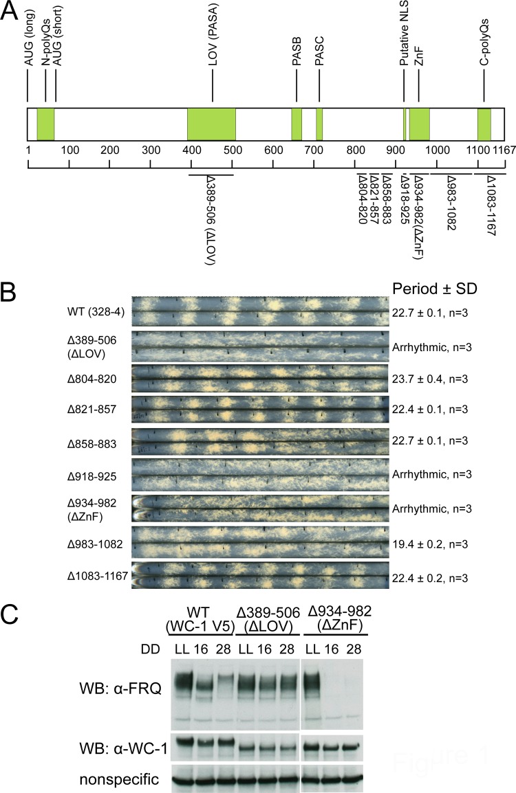
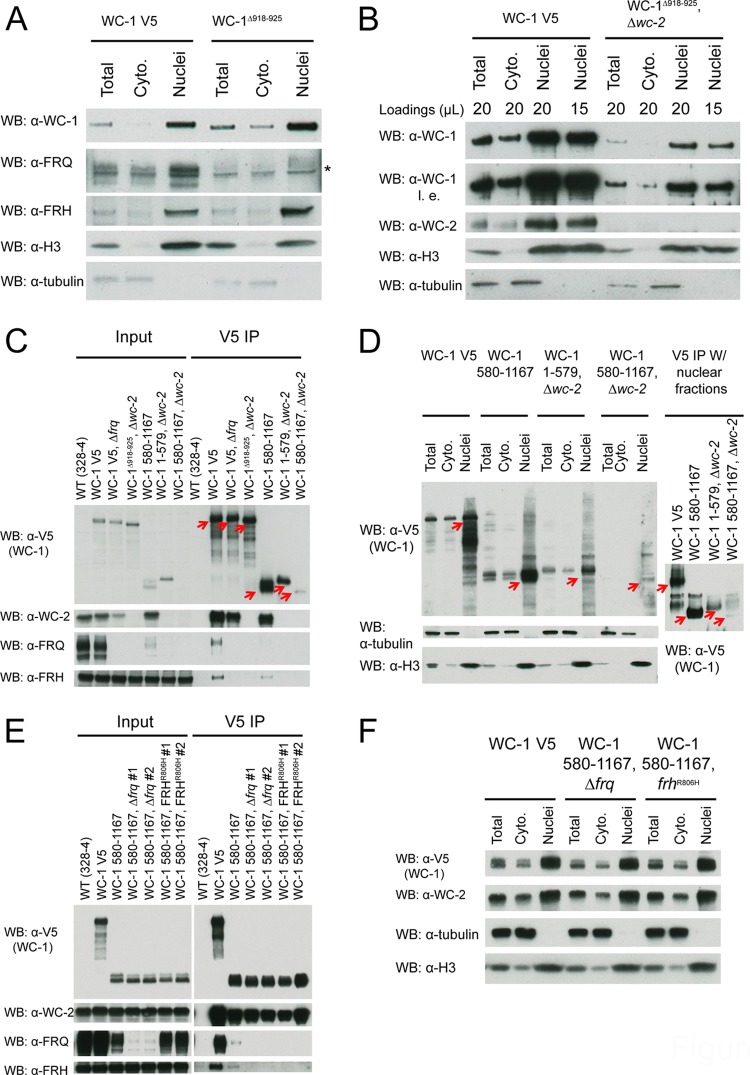
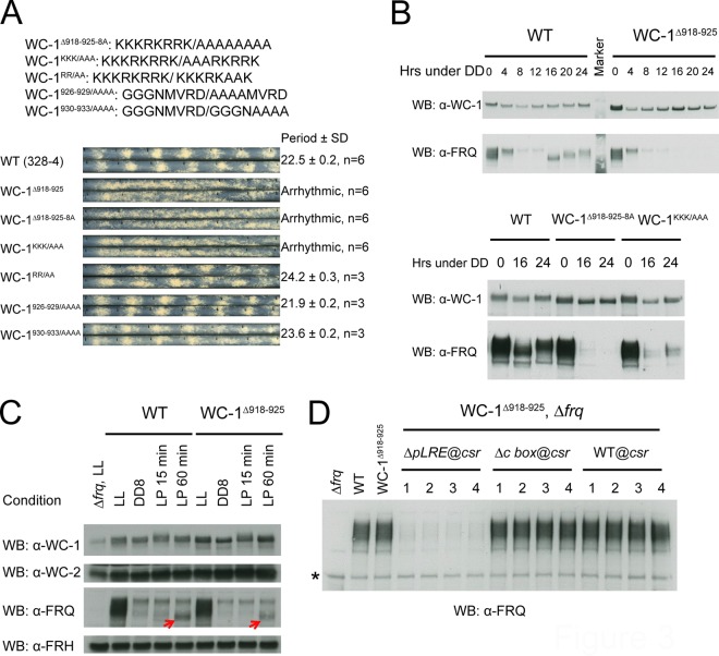
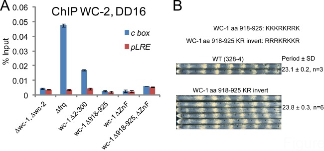
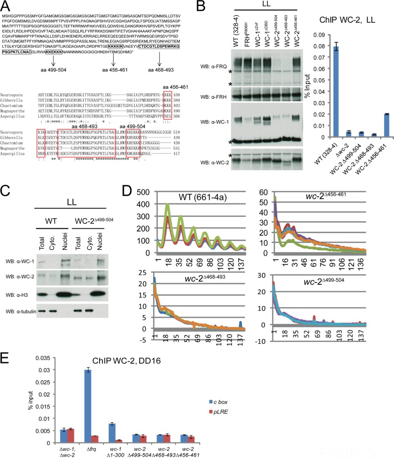
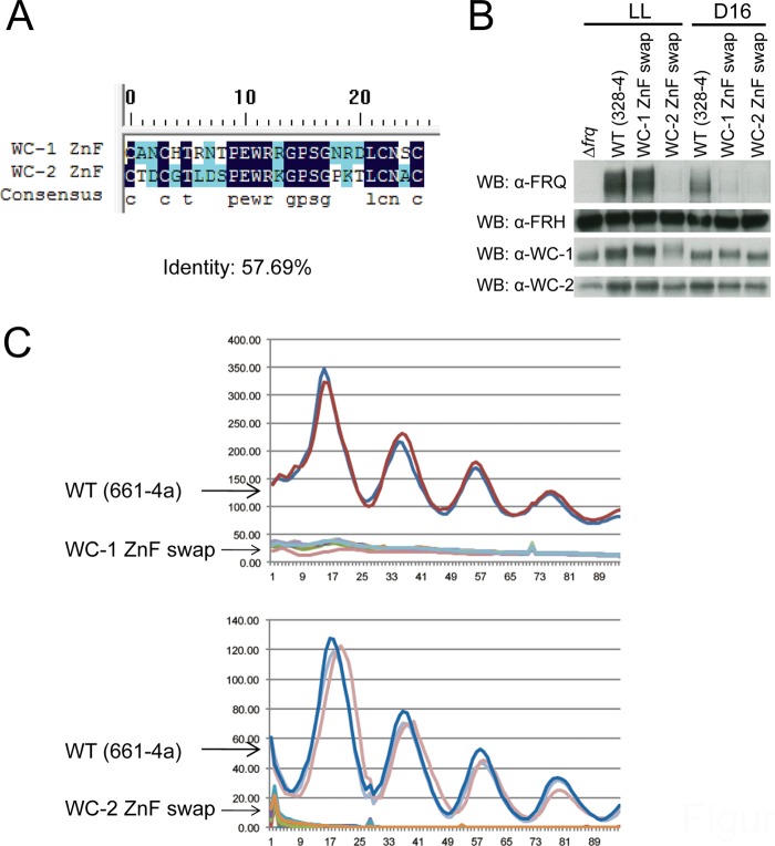
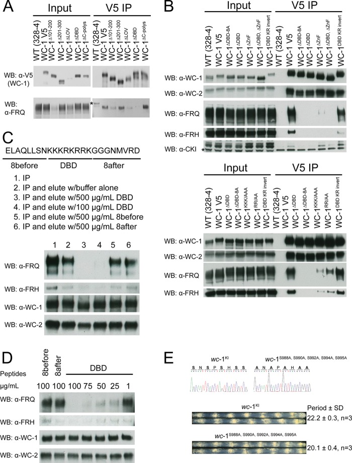
In the Neurospora circadian system, the White Collar complex (WCC) of WC-1 and WC-2 drives transcription of the circadian pacemaker gene frequency (frq), whose gene product, FRQ, as a part of the FRQ-FRH complex (FFC), inhibits its own expression. The WCC is also the principal Neurospora photoreceptor; WCC-mediated light induction of frq resets the clock, and all acute light induction is triggered by WCC binding to promoters of light-induced genes. However, not all acutely light-induced genes are also clock regulated, and conversely, not all clock-regulated direct targets of WCC are light induced; the structural determinants governing the shift from WCC's dark circadian role to its light activation role are poorly described. We report that the DBD region (named for being defective in binding DNA), a basic region in WC-1 proximal to the DNA-binding zinc finger (ZnF) whose function was previously ascribed to nuclear localization, instead plays multiple essential roles assisting in DNA binding and mediating interactions with the FFC. DNA binding for light induction by the WCC requires only WC-2, whereas DNA binding for circadian functions requires WC-2 as well as the ZnF and DBD motif of WC-1. The data suggest a means by which alterations in the tertiary and quaternary structures of the WCC can lead to its distinct functions in the dark and in the light.


Figures

FIG 1
FIG 1
Deletion scanning mutations within WC-1 detect domains of WC-1 required for circadian regulation in Neurospora. (A) Schematic depiction of the domain architecture of the WC-1 protein. Lines with corresponding numbers of amino acids show different deletions of WC-1: ZnF, zinc finger DNA binding domain; NLS, putative nuclear localization signal; LOV, light-, oxygen-, and voltage-sensing domain; PAS, Per-Arnt-Sim domain. (B) Race tube assays of WT (328-4) and WC-1 deletion strains. The strains were inoculated to 0.1% glucose race tube medium and synchronized in the light at 25°C overnight (12 to 16 h) prior to the transition to darkness. Images of replicate race tubes are displayed, and the vertical lines in race tubes mark the growth fronts of the strains every 24 h. Periods were calculated by the ChronOSX program, version 2.1, and are shown at the right in hours (SD, standard deviation; n, number of race tubes). (C) Western blot showing WC-1 and FRQ protein levels in WT and wc-1 mutants. LL, constant light; DD, hours after the light to dark transition. Both WT WC-1 and mutants were C-terminally tagged with V5. DD16 = CT5 (circadian time 5); DD28 = CT17.
FIG 2
FIG 2
WC-1 does not use a tyical basic NLS for import into the nucleus. (A) Western blot analyses showing the enrichment of WC-1Δ918-925 in the nuclear fraction at DD16. Fresh Neurospora tissues were lysed by bead-beating, and nuclei were isolated from cytoplasm by centrifugation and lysed in RIPA buffer as described in Materials and Methods. Tubulin was used as a control to show that the nuclear fractions were not contaminated with cytosolic proteins, while H3 served as a positive control for enrichment of the nuclear fractions. The asterisk indicates a nonspecific band appearing in all fractions. (B) WC-1Δ918-925 was found in the nucleus in the absence of WC-2. Indicated volumes of lysate at the concentration of 1.5 μg/μl were loaded per lane to compare WC-1 levels in different fractions. l. e., longer exposure. (C) Immunoprecipitation assays using V5 antibody showing the levels of WC-1 1-579 and WC-1 580-1167. Arrows point to WC-1 bands. In the absence of WC-2, WC-1 580-1167 is undetected in the input lane (left) but appears as a weak band (right) after enrichment with V5 antibody. For the V5 IP samples, 5 μl out of the 50 μl of total eluate was loaded into each lane. WT (328-4) and WC-1 V5 served as negative and positive controls, respectively, for the IP assay. (D) Western blot analyses showing that in the Δwc-2 background, both WC-1 1-579 and WC-1 580-1167 are located in the nuclear fraction (left). WC-1 580-1167 is also visualized after enrichment with V5 antibody from 2 mg of nuclear proteins (right). Arrows point to WC-1 bands. (E) Immunoprecipitation assays using V5 antibody showing the loss of interaction between WC-1 580-1167 and FRH in the Δfrq mutant or the FFC in the frhR806H mutant. The Neurospora protein lysate was either analyzed directly by Western blotting analyses (input) or immunoprecipitated with V5 antibody (V5 IP). (F) Western blot analyses showing that WC-1 580-1167 in the Δfrq or frhR806H background is located in the nuclear fraction.
FIG 3
FIG 3
The WC-1 basic domain (aa 918 to 925) is essential for operation of the Neurospora clock. (A) Race tube analyses of WC-1Δ918-925 mutants. The identities of the mutations in WC-1 in the region of aa 918 to 925 are listed at the top. The strains were cultured in 0.1% glucose race tube medium in the light at 25°C overnight and transferred to the dark. Vertical lines in race tubes indicate the growth fronts of the strains every 24 h. ChronOSX program version 2.1 was used to calculate circadian periods. WC-1Δ918-925, WC-1Δ918-925-8A, and WC-1Δ918-925-KKK/AAA are arrhythmic at 25°C. (B) Western blot analysis showing that FRQ expression in WC-1Δ918-925 is impaired after transition to the dark (upper blots). In the lower blots, FRQ and WC-1 levels in WC-1Δ918-925-8A and WC-1Δ918-925-KKK/AAA were assayed at DD0 (constant light), DD16 (when new FRQ peaks in the WT), and DD24 (when old FRQ is extensively phosphorylated in the WT). (C) Western blot analysis showing light-induced FRQ expression in WC-1Δ918-925 after a 60-min light exposure. The samples cultured at DD8 were treated with a 15- or 60-min light pulse and immediately harvested and frozen in liquid nitrogen. The arrows point to the FRQ bands. (D) Western blot analysis showing that FRQ expression in WC-1Δ918-925 in response to light requires the pLRE. The wc-1Δ918-925 Δfrq mutant as the recipient strain was transformed with the frq open reading frame (ORF) fused with the C box and pLRE (WT), the pLRE alone (ΔC box), or the C box alone (ΔpLRE) targeting to the csr locus; see Materials and Methods. Expression of FRQ in the light was analyzed by Western blotting. The asterisk indicates a nonspecific band.
FIG 4
FIG 4
The WC-1 DBD motif is required for associating with the C box and for frq transcription in the dark. (A) ChIP at the frq promoter using WC-2 antibody on indicated wc-1 mutants. The Δwc-1 Δwc-2 strain was the negative control, while the Δfrq mutant (in which the frq open reading frame was replaced by the Escherichia coli Hyg resistance-encoding gene [hph]) served as the positive control. Samples at DD16 were formaldehyde cross-linked and harvested by vacuum filtration prior to the addition of SDS lysis buffer and sonication. Quantitative PCR was performed using either the C box- or pLRE-specific oligonucleotides. The bars represent average values plotted as a percentage of the total, with error bars representing the standard errors of the means (SEMs) (n = 3). (B) Race tube assays of WT and WC-1 DBD KR invert strains. Race tubes were prepared as described in Materials and Methods.
FIG 5
FIG 5
WC-2 ZnF (aa 468 to 493) and aa 499 to 504 are required for FRQ expression in the light. (A) Amino acid sequence of WC-2 highlighting aa 456 to 461, the ZnF, and aa 499 to 504, which are in bold and indicated by arrows (upper portion), and alignment of WC-2 ZnF domains from five related fungi (Neurospora crassa, Gibberella zeae, Chaetomium globosum, Magnaporthe grisea, and Aspergillus nidulans) (lower portion). aa 456 to 461, ZnF (aa 468 to 493), and aa 499 to 504 on WC-2s are boxed. WC-2 sequences were downloaded from the NCBI website (http://www.ncbi.nlm.nih.gov/) and aligned using ClustalW2 (http://www.ebi.ac.uk/Tools/msa/clustalw2/). (B) FRQ, FRH, WC-1, and WC-2 levels and ChIP at the frq promoter using WC-2 antibody in the wc-2Δ456-461, wc-2Δ468-493, and wc-2Δ499-504 strains in the light. Asterisks indicate nonspecific protein bands. The bars represent average values taken as a percentage of the total, with error bars showing the SEMs (n = 3). The Δwc-2 strain was the negative control, while the WT (328-4) served as the positive control in the ChIP assay. (C) Western blot analyses showing that WC-2Δ456-461 is located in the nuclear fraction in the light. (D) frq transcription in WT (661-4a), wc-2Δ456-461, wc-2Δ468-493, and wc-2Δ499-504 strains was measured by the frq C box fused with codon-optimized firefly luciferase (transcriptional fusion). Strains cultured in race tube medium bearing luciferin were synchronized by growth in constant light for 2 days, followed by transfer to darkness. The luciferase signal, sampled every 30 min, was tracked for 6 days. Each strain was retested three or more times in the assay. (E) WC-2 DNA binding in the wc-2Δ456-461, wc-2Δ468-493, and wc-2Δ499-504 stains at DD16, when WCC binding to the C box peaks in WT was measured by ChIP assays using WC-2 antibody. The Δwc-1 Δwc-2 strain served as the negative control, while the Δfrq mutant, in which the WCC circadian activity is always on, was the positive control. Quantitative PCR was done using either the C box- or pLRE-specific primer sets (see Materials and Methods). The data are average values plotted as a percentage of the total, with error bars showing the SEMs (n = 3).
FIG 6
FIG 6
Identities of WC-1 and WC-2 ZnFs. (A) Sequence alignment of WC-1 and WC-2 ZnFs by DNAMAN. (B) Western blot analysis of FRQ, FRH, WC-1, and WC-2 levels in the light and at DD16 in the WT, wc-1 ZnF swap, and wc-2 ZnF swap strains. (C) frq transcription was measured using the C box-driven codon-optimized luciferase assays in the wc-1 ZnF swap (top) and wc-2 ZnF swap (bottom) strains. The luciferase signal was taken every 30 min for 4 days. Each strain was retested three times.
FIG 7
FIG 7
The DBD motif in WC-1 is essential for FFC interaction. (A) Immunoprecipitation assays using V5 antibody showing the interaction of WC-1 and FRQ in the WC-1 mutants. The Neurospora protein extracts were either assayed directly with Western blot analyses (left blot) or immunoprecipitated with V5 antibody (right blot). All wc-1 constructs indicated were transformed into the wc-1 native locus of the Δwc-1 strain. WT (328-4) and WC-1 V5 serve as the negative and positive controls, respectively. The immunoprecipitation assays were done as described in Materials and Methods. (B) Immunoprecipitation assays using V5 antibody showing that the DBD motif on WC-1 is required for the FFC-WCC interaction. (C) WCC-FFC interaction can be disrupted by the WC-1 DBD peptide in vitro. The WCC (lane 1) was isolated from WC-1 V5 lysate cultured in the light using V5 antibody as described in Materials and Methods. The washed beads were incubated with protein lysis buffer alone (lane 2), 500 μg/ml of DBD peptide (lane 3), 100 μg/ml of DBD peptide (lane 4), 500 μg/ml of 8before peptide (lane 5), or 500 μg/ml of 8after peptide (lane 6) at 4°C for 1 h, with rotating. After removal of the supernatant, SDS sample buffer was added to the beads prior to elution by boiling, and proteins were assayed by Western blotting analyses. (D) Similar to the case for panel C, WCC-FFC interaction was studied in a titration assay using the WC-1 DBD peptide at concentrations of 100, 75, 50, 25, and 1 μg/ml. (E) Race tube assays of wc-1KI (bearing a WT copy of WC-1) and wc-1S988A, S990A, S992A, S994A, S995A strains (period [in hours] ± 1 standard deviation; n, number of race tubes). Race tube assays were performed as described in Materials and Methods. The five point mutations in the wc-1S988A, S990A, S992A, S994A, S995A strain were confirmed by sequencing genomic DNA as shown in the upper portion.

Similar articles

Cited by

References

    1. Dunlap JC. 1999. Molecular bases for circadian clocks. Cell 96:271–290. doi:10.1016/S0092-8674(00)80566-8. - DOI - PubMed
    1. Mohawk JA, Green CB, Takahashi JS. 2012. Central and peripheral circadian clocks in mammals. Annu Rev Neurosci 35:445–462. doi:10.1146/annurev-neuro-060909-153128. - DOI - PMC - PubMed
    1. Cha J, Zhou M, Liu Y. 2015. Mechanism of the Neurospora circadian clock, a FREQUENCY-centric view. Biochemistry 54:150–156. doi:10.1021/bi5005624. - DOI - PMC - PubMed
    1. Froehlich AC, Loros JJ, Dunlap JC. 2003. Rhythmic binding of a WHITE COLLAR-containing complex to the frequency promoter is inhibited by FREQUENCY. Proc Natl Acad Sci U S A 100:5914–5919. doi:10.1073/pnas.1030057100. - DOI - PMC - PubMed
    1. Froehlich AC, Liu Y, Loros JJ, Dunlap JC. 2002. White Collar-1, a circadian blue light photoreceptor, binding to the frequency promoter. Science 297:815–819. doi:10.1126/science.1073681. - DOI - PubMed

Publication types

MeSH terms

LinkOut - more resources