. 2016 Feb 23;113(8):E1082-8.
doi: 10.1073/pnas.1507109113. Epub 2015 Dec 28.

Pan-neuronal imaging in roaming Caenorhabditis elegans

Affiliations

Pan-neuronal imaging in roaming Caenorhabditis elegans

Vivek Venkatachalam et al. Proc Natl Acad Sci U S A. .

Abstract

We present an imaging system for pan-neuronal recording in crawling Caenorhabditis elegans. A spinning disk confocal microscope, modified for automated tracking of the C. elegans head ganglia, simultaneously records the activity and position of ∼80 neurons that coexpress cytoplasmic calcium indicator GCaMP6s and nuclear localized red fluorescent protein at 10 volumes per second. We developed a behavioral analysis algorithm that maps the movements of the head ganglia to the animal's posture and locomotion. Image registration and analysis software automatically assigns an index to each nucleus and calculates the corresponding calcium signal. Neurons with highly stereotyped positions can be associated with unique indexes and subsequently identified using an atlas of the worm nervous system. To test our system, we analyzed the brainwide activity patterns of moving worms subjected to thermosensory inputs. We demonstrate that our setup is able to uncover representations of sensory input and motor output of individual neurons from brainwide dynamics. Our imaging setup and analysis pipeline should facilitate mapping circuits for sensory to motor transformation in transparent behaving animals such as C. elegans and Drosophila larva.

Keywords: C. elegans; Drosophila; calcium imaging; thermotaxis.


Conflict of interest statement

The authors declare no conflict of interest.

Figures

Fig. 1.
Fig. 1.
Experimental setup. (A) Microscopy schematic. Data were acquired using a spinning disk confocal microscope with red and green channels captured side by side on a CMOS sensor at 200 frames/s. A volume was imaged every 10 s using a piezo objective scanner. Every 20 frames were grouped together to form an image stack. Custom software controlled the stage and objective movements to maintain the tracked target at the center of the image volume. (B) In all experiments, the animal was provided with a sinusoidal temperature stimulus using a custom thermoelectric stage (described in ref. 23). Temperature was recorded using a microthermocouple embedded at the agar surface.
Fig. 2.
Fig. 2.
Image processing for behavior and neuronal activity measurements. (A) We verified the posture reconstruction algorithm by low-magnification (4×) tracking of a worm with the same transgenic line that we used in high-magnification experiments. We used the trajectory of the animal’s nerve ring. The animal was completely visible, but we only used the trajectory of the animal’s brain (information in the yellow box that would be available at 40× magnification) to compute its posture (green lines). The posture qualitatively fits a variety of complex configurations of the crawling worm, even those that are challenging to automatically segment using more conventional image-processing strategies such as omega turns when the head touches the tail. Trajectory represents 221 s of continuous recording starting from (s) and ending at (e). (B) The computer posture and trajectory of an animal recorded at high magnification associated with the pan-neuronal imaging shown in Fig. 3. The algorithm correctly predicts when the worm’s tail will enter the field of view (red posture along the trajectory). Trajectory represents 121 s of continuous recording starting from (s) and ending at (e). (C) By running the algorithm on published behavioral data for N2 worms freely crawling off of food, we can see the goodness of fit as a function of body coordinate (0 at head, 1 at tail). From the nerve ring (0.15 fractional distance from head) to the tail, the algorithm captures most of the variance of body posture. (D) Effectiveness of automated identification of neuronal indexes during brainwide imaging is illustrated by dewarping the nerve ring using our registration algorithm (Methods) that minimizes the displacement of the coordinates of all nuclei from a reference atlas. Three image volumes showing a deep ventral bend, deep dorsal bend, and straight posture from the worm recorded in Fig. 2B are shown. Also see Movies S1 and S2.
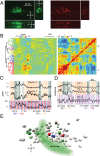
Fig. 3.
Fig. 3.
Multineuronal activity patterns. (A) Images obtained in a 5-ms exposure with our setup in the red channel showing TagRFP and green channel showing cytosolic GCaMP6s. Each image is a slice through the acquired volume. Neurons are resolvable in the green channel, and nuclei are well resolved in the red channel in all dimensions. Signals are attenuated further from the objective. (B) Normalized calcium dynamics of 84 neurons and matrix of correlation coefficients from the trajectory shown in Fig. 2B. Neuronal activity patterns are grouped and ordered by agglomerative hierarchical clustering. Neurons corresponding to the two largest clusters are highlighted in red and green on the tree diagram. Indexes corresponding to the neurons with the strongest signals within each cluster are indicated. (C) Sample traces from a cell activated during forward movement (#7), a cell activated during backward movement (#4), and a cell correlated with temperature oscillations (#32). (D) The first three principal components of the whole-brain neuronal activity correlate most strongly with behavioral output. The first two correlate with velocity, and the third component reports the angular velocity of the head. (E) A volume showing the relative location of segmented neurons in one image volume. Neurons corresponding to the green and red clusters in Fig. 3B are colored accordingly. Neurons discussed in this figure along with Fig. 4 are annotated for reference. Two neurons correlated with thermosensory input are colored in blue. The position of the nerve ring is drawn for reference. The right side of the animal is into the page.
Fig. 4.
Fig. 4.
Stereotyped responses of indexed neurons. (A) High-resolution stacks with simultaneous red (nucleus), green (cytoplasm), and DIC information were used to match the indexes with stereotyped activity patterns to their cellular identities (also see Fig. S1). (B) Consistent signals across animals from temperature-sensitive neurons (32-AFDL and 69-AFDR) and several neurons correlated with forward/backward movement, suggesting that the indexes assigned using DIC are the same as those recorded in roaming animals. Four worms (–4) represent our standard imaging conditions with pan-neuronal GCaMP6s. The GFP control worm was subjected to the same imaging and analysis procedure, but using a transgenic worm that expressed pan-neuronal GFP. The residual signal in the control worm provides an estimate of total noise in the system caused by experimental measurement and the analysis pipeline.

Comment in

  • Grabbing brain activity on the go.
    Clandinin TR, Goodman MB. Clandinin TR, et al. Proc Natl Acad Sci U S A. 2016 Feb 23;113(8):1965-7. doi: 10.1073/pnas.1524219113. Epub 2016 Feb 3. Proc Natl Acad Sci U S A. 2016. PMID: 26842834 Free PMC article. No abstract available.

References

    1. Faumont S, et al. An image-free opto-mechanical system for creating virtual environments and imaging neuronal activity in freely moving Caenorhabditis elegans. PLoS One. 2011;6(9):e24666. - PMC - PubMed
    1. Kawano T, et al. An imbalancing act: Gap junctions reduce the backward motor circuit activity to bias C. elegans for forward locomotion. Neuron. 2011;72(4):572–586. - PubMed
    1. Clark DA, Gabel CV, Gabel H, Samuel AD. Temporal activity patterns in thermosensory neurons of freely moving Caenorhabditis elegans encode spatial thermal gradients. J Neurosci. 2007;27(23):6083–6090. - PMC - PubMed
    1. Suzuki H, et al. In vivo imaging of C. elegans mechanosensory neurons demonstrates a specific role for the MEC-4 channel in the process of gentle touch sensation. Neuron. 2003;39(6):1005–1017. - PubMed
    1. Chalasani SH, et al. Dissecting a circuit for olfactory behaviour in Caenorhabditis elegans. Nature. 2007;450(7166):63–70. - PubMed

Publication types

Substances