. 2016 Jan 12;113(2):374-9.
doi: 10.1073/pnas.1519691113. Epub 2015 Dec 28.

A second transmissible cancer in Tasmanian devils

Affiliations

A second transmissible cancer in Tasmanian devils

Ruth J Pye et al. Proc Natl Acad Sci U S A. .

Abstract

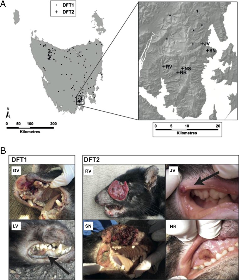
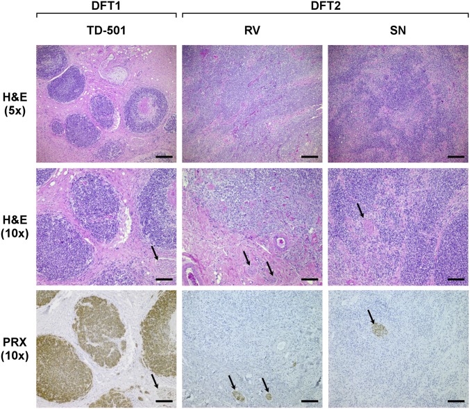
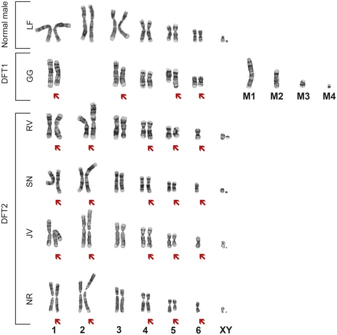
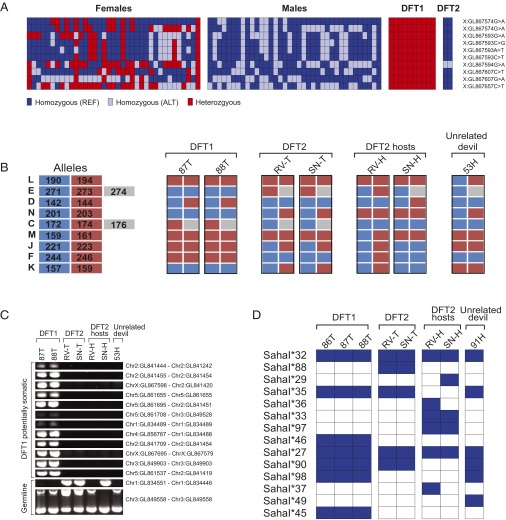
Clonally transmissible cancers are somatic cell lineages that are spread between individuals via the transfer of living cancer cells. There are only three known naturally occurring transmissible cancers, and these affect dogs, soft-shell clams, and Tasmanian devils, respectively. The Tasmanian devil transmissible facial cancer was first observed in 1996, and is threatening its host species with extinction. Until now, this disease has been consistently associated with a single aneuploid cancer cell lineage that we refer to as DFT1. Here we describe a second transmissible cancer, DFT2, in five devils located in southern Tasmania in 2014 and 2015. DFT2 causes facial tumors that are grossly indistinguishable but histologically distinct from those caused by DFT1. DFT2 bears no detectable cytogenetic similarity to DFT1 and carries a Y chromosome, which contrasts with the female origin of DFT1. DFT2 shows different alleles to both its hosts and DFT1 at microsatellite, structural variant, and major histocompatibility complex (MHC) loci, confirming that it is a second cancer that can be transmitted between devils as an allogeneic, MHC-discordant graft. These findings indicate that Tasmanian devils have spawned at least two distinct transmissible cancer lineages and suggest that transmissible cancers may arise more frequently in nature than previously considered. The discovery of DFT2 presents important challenges for the conservation of Tasmanian devils and raises the possibility that this species is particularly prone to the emergence of transmissible cancers. More generally, our findings highlight the potential for cancer cells to depart from their hosts and become dangerous transmissible pathogens.

Keywords: Tasmanian devil; Tasmanian devil facial tumor disease; contagious cancer; transmissible cancer.


Conflict of interest statement

The authors declare no conflict of interest.

Figures

Fig. 1.
Fig. 1.
Geographical location and gross appearance of DFT2 tumors. (A) Locations of confirmed DFT1 and DFT2 tumors in Tasmania (Left) and the Channel Peninsula (Right). Each DFT1 location is represented with a single dot regardless of the number of tumors identified at this location. Tumor diagnosis was performed by histopathology, cytogenetics, and/or genetic analysis. (B) Gross appearance of two DFT1 tumors (Left) and four DFT2 tumors (Right). Tumors were identified in Tasmanian devils in the Channel region between 2012 and 2015. Further information about animals is available in Table S1.
Fig. 2.
Fig. 2.
DFT2 tumors are histologically distinct from DFT1. Representative images of H&E stained histological sections of DFT1 and DFT2 tumors (Upper and Middle). (Lower) Histological sections stained with DFT1 marker, PRX. Scale bars represent 200 μm (Upper) or 100 μm (Middle and Lower). Arrows indicate peripheral nerve bundles, which are positive for PRX.
Fig. 3.
Fig. 3.
DFT2 tumors are cytogenetically distinct from DFT1. Representative karyotypes of a normal male devil, a DFT1 tumor, and four DFT2 tumors. Red arrows indicate chromosomes carrying cytogenetic abnormalities. Four marker chromosomes found in DFT1 (9) are labeled M1 to M4. Karyotype for NS is presented in Fig. S1.
Fig. S1.
Fig. S1.
Representative karyotype for DFT2 tumor derived from animal NS.
Fig. 4.
Fig. 4.
DFT2 tumors are genetically distinct from DFT1 and from their hosts. (A) Genotyping of X-linked SNPs. Genotypes of 37 female and 37 male devils at 10 X-linked SNP loci that are heterozygous in DFT1. Both homozygous and hemizygous genotypes are referred to as homozygous. Each individual is represented by a column, and chromosome and scaffold for each locus is shown. Further information about individuals and exact SNP coordinates are available in Tables S3 and S4. (B) Microsatellite genotypes at nine polymorphic microsatellite loci (L, E, D, N, C, M, J, F, and K). The lengths of the two (L, D, N, M, J, F, and K) or three (E and C) alleles found at each locus and their color codes are shown on the left, and their genotypes in DFT1 tumors, DFT2 tumors, DFT2 hosts, and in 53H, a representative unrelated devil, are shown. All tumors are presented as diploid, although true copy number at these loci is not known. Further information about allele sizes is found in Table S2. (C) Structural variant genotyping. PCRs spanning breakpoint junctions were performed to assess the presence or absence of twelve possibly somatic structural variants and two polymorphic germ-line variants. We cannot confirm if structural variants found only in DFT1 are somatic or rare germ-line variants, thus these are labeled potentially somatic. Chromosomes and scaffolds involved in each rearrangement are indicated, and full breakpoint coordinates are available in Table S5. (D) MHC class I exon 2 haplotypes detected in DFT1 tumors, DFT2 tumors, DFT2 hosts, and in 91H, a representative unrelated devil. Exon 2 haplotype names are indicated on the left, and their presence or absence in the panel of samples are indicated with blue and white squares respectively. All 14 haplotypes are predicted to encode a unique amino acid sequence. Complete sequences for each haplotype are available in Tables S6 and S7.

References

    1. Metzger MJ, Reinisch C, Sherry J, Goff SP. Horizontal transmission of clonal cancer cells causes leukemia in soft-shell clams. Cell. 2015;161(2):255–263. - PMC - PubMed
    1. Murchison EP. Clonally transmissible cancers in dogs and Tasmanian devils. Oncogene. 2008;27(Suppl 2):S19–S30. - PubMed
    1. Strakova A, Murchison EP. The cancer which survived: Insights from the genome of an 11000 year-old cancer. Curr Opin Genet Dev. 2015;30:49–55. - PubMed
    1. Hawkins CE, et al. Emerging disease and population decline of an island endemic, the Tasmanian devil Sarcophilus harrisii. Biol Conserv. 2006;131:307–324.
    1. Loh R, et al. The pathology of devil facial tumor disease (DFTD) in Tasmanian Devils (Sarcophilus harrisii) Vet Pathol. 2006;43(6):890–895. - PubMed

Publication types

Associated data