. 2016 Feb 2;113(5):E626-34.
doi: 10.1073/pnas.1522821113. Epub 2015 Dec 28.

GR SUMOylation and formation of an SUMO-SMRT/NCoR1-HDAC3 repressing complex is mandatory for GC-induced IR nGRE-mediated transrepression

Affiliations

GR SUMOylation and formation of an SUMO-SMRT/NCoR1-HDAC3 repressing complex is mandatory for GC-induced IR nGRE-mediated transrepression

Guoqiang Hua et al. Proc Natl Acad Sci U S A. .

Abstract

Unique among the nuclear receptor superfamily, the glucocorticoid (GC) receptor (GR) can exert three distinct transcriptional regulatory functions on binding of a single natural (cortisol in human and corticosterone in mice) and synthetic [e.g., dexamethasone (Dex)] hormone. The molecular mechanisms underlying GC-induced positive GC response element [(+)GRE]-mediated activation of transcription are partially understood. In contrast, these mechanisms remain elusive for GC-induced evolutionary conserved inverted repeated negative GC response element (IR nGRE)-mediated direct transrepression and for tethered indirect transrepression that is mediated by DNA-bound NF-κB/activator protein 1 (AP1)/STAT3 activators and instrumental in GC-induced anti-inflammatory activity. We demonstrate here that SUMOylation of lysine K293 (mouse K310) located within an evolutionary conserved sequence in the human GR N-terminal domain allows the formation of a GR-small ubiquitin-related modifiers (SUMOs)-NCoR1/SMRT-HDAC3 repressing complex mandatory for GC-induced IR nGRE-mediated direct repression in vitro, but does not affect transactivation. Importantly, these results were validated in vivo: in K310R mutant mice and in mice ablated selectively for nuclear receptor corepressor 1 (NCoR1)/silencing mediator for retinoid or thyroid-hormone receptors (SMRT) corepressors in skin keratinocytes, Dex-induced direct repression and the formation of repressing complexes on IR nGREs were impaired, whereas transactivation was unaffected. In mice selectively ablated for histone deacetylase 3 (HDAC3) in skin keratinocytes, GC-induced direct repression, but not bindings of GR and of corepressors NCoR1/SMRT, was abolished, indicating that HDAC3 is instrumental in IR nGRE-mediated repression. Moreover, we demonstrate that the binding of HDAC3 to IR nGREs in vivo is mediated through interaction with SMRT/NCoR1. We also show that the GR ligand binding domain (LBD) is not required for SMRT-mediated repression, which can be mediated by a LBD-truncated GR, whereas it is mandatory for NCoR1-mediated repression through an interaction with K579 in the LBD.

Keywords: GC-induced direct transrepression; IR nGRE; SUMOylation; glucocorticoid receptor.


Conflict of interest statement

The authors declare no conflict of interest.

Figures
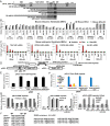
Fig. 1.
Fig. 1.
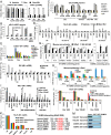
GR binding to an IR nGRE in vitro and in vivo requires a discrete sequence located in the NTD. (A) GR NTD and LBD deletion mutants. (B) Quantitative (q)RT-PCR for IR nGRE-containing genes in GRwt and GRα-D3 MEFs. Vehicle, Dex (0.5 µM), and RU (3 µM) treatments were for 6 h. (C) qPCR of ChIP assays performed with GRwt and GRα-D3 MEFs treated with vehicle, Dex (1 µM), and RU (6 µM) for 1 h, showing the association of GR, nuclear receptor corepressor 1, and silencing mediator for retinoid or thyroid-hormone receptors (SMRT) on IR nGREs in the promoter region of genes as indicated. (D) Schematic representation for pGL3-17mer Gal4 reporter, pSG5-Gal4, and pSG5-GR NTD-Gal4 plasmids. (E) Luciferase assays of Cos-1 cells transfected with pGL3-17mer Gal4 reporter and pSG5-Gal4 alone or fused to GR NTD or its mutants as indicated (see D). (F) As in C, but performed with Cos-1 cells transfected with pGL3-17mer Gal4 reporter and pSG5-Gal4 constructs (see D), showing the association of indicated proteins on the Gal4 RE sequence, using specific antibodies. (G) As in E, but transfected with pGL3-nGRE reporter and GR mutants. Vehicle or Dex (0.5 µM) treatment was for 6 h. (H and I) As in F but transfected with pGL3-nGRE reporter and GR mutants, showing the binding of GR, NCoR1, and SMRT on an IR nGRE. Cells were treated with vehicle or Dex (1 µM) for 1 h. (J) Alignment of the 283–295 AA sequence in human GR with homologous GR sequences in vertebrates (Left) and predicted SUMOylation sites in the human and mouse GR sequence (Right). Values are mean ± SEM. *P < 0.05, **P < 0.01.
Fig. S1.
Fig. S1.
GR binding to an IR nGRE in vitro requires a discrete sequence located in the NTD. (A) Immunoblots of WCE from Cos-1 cells transfected with GR and NTD mutants, NT, not transfected; arrow, nonspecific protein. (B) Schematic representation of pGL3 reporter. (C) Immunoblots of NE from Cos-1 cells transfected with GR or GR ABCD, treated with vehicle, Dex, or RU486 (0.5 µM) for 1 h. Sp1 factor was a control for nuclear localization. (D and G) Luciferase assays of Cos-1 cells transfected with pGL3 luciferase reporters and GR NTD mutants. Vehicle or Dex (0.5 µM) treatment was for 6 h. (E and H) qRT-PCR for SGK1 and STRA13 transcripts in transfected Cos-1 cells. Vehicle, Dex (0.5 µM) and RU (3 µM) treatments were for 6 h. (F and I) qPCR of ChIP assays performed with Cos-1 cells transfected with pGL3 reporters and GR NTD mutants, using GR antibody to show GR binding on a (+)GRE or an IR nGRE. Cells were treated with vehicle or Dex (1 µM) for 1 h. IgG control for IP was ∼0.02% of input. (J) Luciferase assays of Cos-1 cells transfected with pGL3-nGRE reporter, GR ABCD, or GR K579A and indicated siRNA. Vehicle or Dex (0.5 µM) treatment was for 6 h. (K and L) qPCR analyses of ChIP assays performed with Cos-1 cells transfected as in J, showing the binding of indicated proteins to IR1 nGRE, using specific antibodies. Cells were treated with vehicle or Dex (1 µM) for 1 h. Values are mean ± SEM. *P < 0.05.
Fig. S2.
Fig. S2.
Generation of GR mutant mice selectively expressing GRα-D3 isoform. (A) Strategy of the insertion of LoxP sites into the first exon of murine GR genomic locus. (B) Schematic representation of conditional deletion of 352 AAs from the GR NTD. The primers indicated are used for GR transcript assessment. (C) Immunoblot analyses for GR expression using liver samples from E13 WT, GRF/F, and GRF/+ embryos. GAPDH was used as a loading control. (D) Immunoblot analyses for GR expression using WT (lane 3) and GRα-D3 (lane 2) mice liver samples. Lysates from Cos-1 cell transfected with either GR FL (lane 4) or GR 336 (lane 1) were used as controls. Arrows indicated the bands for GR WT, GRα-D3, and nonspecific protein. (E) qRT-PCR for transcripts of GR expression using liver samples from WT, GRF/F and GRF/+ embryos, or WT and GRα-D3 mice; primers as indicated in B. Values are mean ± SEM.
Fig. 2.
Fig. 2.
GR SUMOylation at K293 in the GR NTD is crucial for Dex-induced IR nGRE-mediated direct transrepression through interaction between SUMOs and corepressors NCoR1 and SMRT. (A) qRT-PCR for STRA13 and serum and glucocorticoid-regulated kinase 1 (SGK1) transcripts in Cos-1 cells transfected with GR bearing mutations in SUMOylation sites, treated with vehicle, Dex (0.5 µM), and RU (3 µM) for 6 h. (B and C) qPCR of ChIP assays using Cos-1 cells transfected with pGL3-nGRE reporter and GR expression vectors, treated with vehicle, Dex (1 µM), or RU (1 or 6 µM for the cotreatment) for 1 h, showing the binding of GR, NCoR1, SMRT, SUMO1, and SUMO2/3 to an IR nGRE sequence. (D) As in B, but showing the association of indicated proteins on the IR1 nGRE of the TSLP gene. (E) As in D, but using WT mouse dorsal epidermis topically treated with vehicle, Dex (6 nmol/cm2) and RU (36 nmol/cm2) for 6 h. (F) As in B, but on the IR1 nGRE present in exon 6 of the GR gene. (G) As in A, but for IR nGRE and (+)GRE-containing genes from ear extracts of GRwt and GR K310R mutant mice, treated with vehicle or Dex (6 nmol/cm2) for 18 h. (H) As in E, but with dorsal epidermis from WT and GR K310R mutant mice topically treated with Dex, showing the association of GR, SUMO1, and SMRT on indicated DBSs. (I) As in D, but using Cos-1 cells transfected with GR, GR S226A/S404A, or GR 5SA, treated as indicated. (J) As in D, but using A549 cells treated as indicated; 25 µM JNK inhibitor II or/and 20 mM LiCl was added into the medium 30 min before Dex treatment. (K) As in I, but transfected with GR ABCD or its mutants. (L) Sequence alignment of DAXX SIM with putative human and mouse NCoR1 and SMRT SIM. (M) GST-pull down of 35S-labeled C-terminal moieties of NCoR1 and SMRT proteins by GST or GST-SUMO1 protein. Input, 10% of 35S-labeled proteins used for the binding assay. Values are mean ± SEM. *P < 0.05, **P < 0.01.
Fig. S3.
Fig. S3.
GR SUMOylation at K293 in the GR NTD is crucial for Dex-induced IR nGRE-mediated direct transrepression. (A and C) Luciferase assays of Cos-1 cells transfected with pGL3-nGRE or pGL3-(+)GRE reporters and GR or GR ABCD bearing mutations in SUMOylation sites, treated with vehicle or Dex (0.5 µM) for 6 h. (B) SUMOylation assays performed in Cos-1 cells transfected with GR and SUMO1, treated with or without 1 µM Dex for 1 h. WCEs were immunoprecipitated with a GR antibody and washed five times in RIPA buffer. Eluates were electrophoresed and immunoblotted against GR antibody. (D and E) qPCR analyses of ChIP assays performed with Cos-1 cells transfected with pGL3-(+)GRE reporter and GR mutants as indicated, using specific antibodies to show the bindings of GR, NCoR1, SMRT, and SUMOs on a (+)nGRE. Cells were treated with vehicle or Dex (1 µM) for 1 h. (F) qRT-PCR for transcripts from IR nGRE-containing genes TSLP and STRA13 in A549 cells transfected with or without DAXX expression vector, and treated with vehicle, 0.5 µM Dex, and 3 µM RU for 6 h. (G) As in C, but using Cos-1 cells transfected with GR ± DAXX, treated with vehicle, Dex (1 µM), and RU (6 µM) for 1 h, showing the binding of indicated proteins on the IR nGREs of the TSLP and STRA13 genes. Values are mean ± SEM.
Fig. 3.
Fig. 3.
NCoR1 and/or SMRT corepressors and HDAC3 are required for Dex-induced IR nGRE-mediated transrepression in vivo. (A) qRT-PCR for IR nGRE-containing genes in MEFs derived from WT, NCoR1−/−, or SMRT−/− mouse embryos, treated with vehicle, Dex (0.5 µM), and RU (3 µM) for 6 h. (B) qPCR analyses of ChIP assays performed with WT, SMRT−/−, or NCoR1−/− MEFs, treated with vehicle, Dex (1 µM), and RU (6 µM) for 1 h, showing GR and corepressors bindings to the indicated IR nGRE regions. (C) As in A, but using mouse ear epidermis in WT, SMRTep−/− and/or NCoR1ep−/− mutant mice. Mouse ears were treated with vehicle, Dex (6 nmol/cm2), and RU (36 nmol/cm2) for 18 h. (D) As in B, but using dorsal epidermis of SMRTep−/− and/or NCoR1ep−/− mutant mice, treated as in C for 6 h. (E) As in C, but in WT and HDAC3ep−/− mutant mice. (F) As in D, but in WT and HDAC3ep−/− mutant mice. Values are mean ± SEM.
Fig. S4.
Fig. S4.
NCoR1 and/or SMRT corepressors and HDAC3 are required for Dex-induced IR nGRE-mediated repression in vivo. (A) qRT-PCR for SMRT and NCoR1 transcripts in MEFs derived from WT, NCoR1−/−, or SMRT−/− mouse embryos. (B) As in A, but for IR nGRE-containing genes in MEFs, treated with vehicle, Dex (0.5 µM), and RU (3 µM) for 6 h. (C) qPCR analyses of ChIP assays performed with WT, SMRT−/−, or NCoR1−/− MEFs, treated with vehicle, Dex (1 µM), and RU (6 µM) for 1 h, showing GR and corepressors bindings to the indicated IR nGRE regions. (D) As in A, but from ear epidermis in WT, SMRTep−/−, and/or NCoR1ep−/− mutant mice. (E) As in D but for (+)GRE-containing genes from [SMRT/NCoR1]ep−/− mutant mice. Mouse ears were treated with vehicle, Dex (6 nmol/cm2), and RU (36 nmol/cm2) for 18 h. (F) As in C, but using dorsal epidermis from [SMRT/NCoR1]ep−/− mutant mice, treated as in E for 6 h showing the binding of indicated proteins to the [(+)GRE]2x region of FKBP5 gene. (G) As in D, but for HDAC3 transcripts from mouse ear epidermis in WT and HDAC3ep−/− mutant mice. Values are mean ± SEM.
Fig. S5.
Fig. S5.
SUMOylation of the GR did not affect (+)GRE-mediated transactivation. (A) qPCR analyses of ChIP assays performed with Cos-1 cells transfected with GR or GR K293R, treated with vehicle, Dex (1 µM), and RU (6 µM) for 1 h, showing the association of indicated proteins on the (+)GRE region of genes as indicated. (B) As in A, but for IR nGRE regions.
Fig. 4.
Fig. 4.
SUMOylation of the GR did not affect (+)GRE-mediated transactivation. (A) Schematic representation for pGL3 luciferase reporters containing either a single (+)GRE or a [(+)GRE]3× DBS together with an IR nGRE DBS. (B) qPCR analyses of ChIP assays performed with Cos-1 cells transfected with GR and luciferase reporters (as in A), treated with vehicle, Dex (1 µM), and RU (6 µM) for 1 h, showing the binding of GR, cofactors, and SUMOs on indicated regions.
Fig. 5.
Fig. 5.
The GR DBD is differently involved in (+)GRE-mediated transactivation and IR nGRE-mediated transrepression. (A) Relative RNA transcripts (normalized to GR full length, taken as 100%) for GR mutants as indicated (Fig. S6 A and B). (B) As in A, but for the binding of GR mutants to DBS of indicated genes (Fig. S6 A and C). (C) Representation of interactions between GR DBD AAs and TSLP IR1 nGRE bases and between GR DBD AAs and consensus (+)GRE bases. (D) qPCR analyses of ChIP assays performed with A549 cells, treated with vehicle, IL6 (10 ng/mL), and Dex (1 µM) for 1 h, showing the binding of GR, STAT3, and corepressors to the IR0 nGRE and SBE of the SOCS3 gene. Values are mean ± SEM.
Fig. S6.
Fig. S6.
The GR DBD is differently involved in (+)GRE-mediated transactivation and IR nGRE-mediated transrepression. (A) Location of point mutations within the zinc fingers (ZF1 and ZF2) of human GR. Residues that prevent the GR from interacting with (+)GRE and IR nGRE are indicated by black dot and red star, respectively. (B) qRT-PCR for transcripts from the SGK1 and STRA13 genes in Cos-1 cells transfected with GR or GR DBD mutants, treated with vehicle, 0.5 µM Dex, and 3 µM RU for 6 h. (C) qPCR analyses of ChIP assays performed with Cos-1 cells transfected with GR or GR DBD mutants, treated with vehicle or 1µM Dex for 1 h, showing the binding of GR to the SGK1 (+)GRE and TSLP IR1 nGRE, using GR antibody. The IgG control for i.p. (∼0.02% of input) is not shown. (D) IR nGRE sequences present in the indicated genes. Values are mean ± SEM. **P < 0.01.

Comment in

  • A trilogy of glucocorticoid receptor actions.
    Tan CK, Wahli W. Tan CK, et al. Proc Natl Acad Sci U S A. 2016 Feb 2;113(5):1115-7. doi: 10.1073/pnas.1524215113. Epub 2016 Jan 20. Proc Natl Acad Sci U S A. 2016. PMID: 26792523 Free PMC article. No abstract available.

Similar articles

Cited by

References

    1. Clark AR, Belvisi MG. Maps and legends: The quest for dissociated ligands of the glucocorticoid receptor. Pharmacol Ther. 2012;134(1):54–67. - PubMed
    1. Cruz-Topete D, Cidlowski JA. One hormone, two actions: Anti- and pro-inflammatory effects of glucocorticoids. Neuroimmunomodulation. 2015;22(1-2):20–32. - PMC - PubMed
    1. Meijsing SH, et al. DNA binding site sequence directs glucocorticoid receptor structure and activity. Science. 2009;324(5925):407–410. - PMC - PubMed
    1. Ratman D, et al. How glucocorticoid receptors modulate the activity of other transcription factors: A scope beyond tethering. Mol Cell Endocrinol. 2013;380(1-2):41–54. - PubMed
    1. Vayssière BM, et al. Synthetic glucocorticoids that dissociate transactivation and AP-1 transrepression exhibit antiinflammatory activity in vivo. Mol Endocrinol. 1997;11(9):1245–1255. - PubMed

Publication types

MeSH terms