. 2016 Jun;125(2):253-64.
doi: 10.1007/s00412-015-0564-3. Epub 2015 Dec 28.

E-type cyclins modulate telomere integrity in mammalian male meiosis

Affiliations

E-type cyclins modulate telomere integrity in mammalian male meiosis

Marcia Manterola et al. Chromosoma. 2016 Jun.

Abstract

We have shown that E-type cyclins are key regulators of mammalian male meiosis. Depletion of cyclin E2 reduced fertility in male mice due to meiotic defects, involving abnormal pairing and synapsis, unrepaired DNA, and loss of telomere structure. These defects were exacerbated by additional loss of cyclin E1, and complete absence of both E-type cyclins produces a meiotic catastrophe. Here, we investigated the involvement of E-type cyclins in maintaining telomere integrity in male meiosis. Spermatocytes lacking cyclin E2 and one E1 allele (E1+/-E2-/-) displayed a high rate of telomere abnormalities but can progress to pachytene and diplotene stages. We show that their telomeres exhibited an aberrant DNA damage repair response during pachynema and that the shelterin complex proteins TRF2 and RAP2 were significantly decreased in the proximal telomeres. Moreover, the insufficient level of these proteins correlated with an increase of γ-H2AX foci in the affected telomeres and resulted in telomere associations involving TRF1 and telomere detachment in later prophase-I stages. These results suggest that E-type cyclins are key modulators of telomere integrity during meiosis by, at least in part, maintaining the balance of shelterin complex proteins, and uncover a novel role of E-type cyclins in regulating chromosome structure during male meiosis.

Keywords: Cyclin E1; Cyclin E2; E-type cyclins; Meiosis; Meiosis control; Shelterin complex; TRF1; TRF2; Telomere; Telomere integrity.


Figures

Figure 1
Figure 1. Deficiency of E-type cyclins produces abnormal DNA damage at the ends of the chromosomes, preferentially at the proximal end
(A) Localization of γH2AX (green), centromeric proteins (CEN) (white), and SYCP3 (red) during the pachytene stage in wild type (WT) (a-b) and E1+/− E2−/− (c-d) spermatocyte spread preparations. XY indicates the sex body. Enlargements to the upper right of each panel display the chromosomes demarcated by the white rectangle in the full image. The bottom panel corresponds to chromosomes shown above but only γH2AX and CEN localization is depicted, to facilitate the visualization of the γH2AX foci. (a,c) Early pachytene spermatocytes. (a) γH2AX localizes in the sex chromosomes (XY) but also in the chromatin close to the SC in synapsed regions (enlargement). (c) In E1+/− E2−/− spermatocytes, γH2AX also localizes at chromosome ends (yellow arrows, enlargement) as well as in other chromosome regions. (b,d) Mid pachytene spermatocytes. (b) In WT autosomes, very few γH2AX foci persist in the chromatin close to the SC. (d) In E1+/− E2−/− autosomes, γH2AX foci persist in the chromatin, mainly at the chromosome ends and notably in the proximal ends (yellow arrows, enlargement). (B) Scheme for localizing and quantifying the distribution of γH2AX foci. Top panel: example of an early pachytene WT chromosome (demarcated by the white rectangle in panel A(a) immunolocalized for γH2AX (green), CEN (white), and SYCP3 (red). The bottom panel corresponds to the chromosome shown above but only γH2AX and CEN localization is depicted. Five discrete equal segments were defined along the chromosome length. The first and last segments were divided in two sub-segments of equal length and named proximal (p) (yellow) and sub-proximal (s-p) (orange), if they were in or next to the centromere; and sub-distal (s-d) (blue) and distal (d) (green), if they were in the opposite end of the chromosome from the centromere. The other three segments were designated central (c) (white). These segments were used to define the distribution of γH2AX foci along the chromosome length. (C) Distribution of the γH2AX foci along the chromosome segment per spermatocyte, displayed as a percentage of the total number of segments (defined in B) from WT (black bars) and E1+/− E2−/− (dotted bars) chromosomes. Four spermatocytes each from early, mid and late pachynema from 3 different animals were analyzed and quantified (total n= 36). Error bars indicate standard deviation. * p< 0.05, ** p< 0.01, *** p< 0.001.
Figure 2
Figure 2. γH2AX foci co-localize with TRF1 at the telomeres in E1+/−E2−/− pachytene spermatocytes, although the levels of TRF1 are normal
(A) Immunolocalization of γH2AX (blue), TRF1 (red) and SYCP3 (grey) in squash preparations of pachytene spermatocytes. (a,b) Confocal images of WT (a) and E1+/− E2−/− (b) mid pachytene nuclei. XY indicates the sex body. Small panels to the upper right of the main figures enlarge the ends of chromosomes positive for TRF1 localization, demarcated by the white rectangles in a and b. Lower small panels correspond to the chromosomes ends shown above, but only γH2AX and TRF1 localization is depicted. (a) WT nucleus. All telomeres are devoid of γH2AX. (b) In cyclin E-deficient nuclei, a subset of telomeres exhibit γH2AX signal co-localizing with TRF1 in the nuclear periphery (yellow arrows, enlargement). (B) TRF1 (green), centromeric proteins (CEN) (white) and SYCP3 (red) in mid pachytene chromosome spreads of WT and E1+/− E2−/− spermatocytes. XY indicates the sex chromosomes. As above, upper panels enlarge the chromosomes demarcated by the white rectangle in (a) and (b). Lower panels correspond to chromosomes shown above but only TRF1 and CEN localization is depicted. (a) WT mid pachytene spermatocyte with defined TRF1 signal in all telomeres (enlargement). (b) E1+/− E2−/− mid pachytene spermatocyte exhibit telomeres with TRF1 in the telomeres (enlargements). In some chromosomes, TRF1 localizes forming bridges between two or more chromosomes (white arrow) or fused telomeres (blue arrow). (C) Quantification of the intensity of TRF1 signal in proximal and distal telomeres in both WT (black bars) and E1+/− E2−/− (dotted bars)chromosomes. n= 36 spermatocytes. Error bars indicate standard deviation.
Figure 3
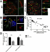
Figure 3. The presence of γH2AX foci at the telomeres is correlated with reduced levels of TRF2, mainly in the proximal telomeres
(A) Localization of TRF2 (green), centromeric proteins (CEN) (white) and SYCP3 (red) in mid pachytene spermatocyte spread preparations. Small panels to the right of the main image highlight the chromosomes demarcated by white rectangles in (a) and (b). Upper panel of each set shows localization of SYCP3 (red), TRF2 (green), and CEN (white) while the lower panels depict only CEN and TRF2 signal. XY indicates the sex chromosomes. (a) WT pachytene spermatocyte with defined TRF2 signal in all telomeres. (b) E1+/− E2−/− spermatocyte with reduced signal of TRF2 at the telomeres. Yellow arrows indicate telomeres with significantly reduced TRF2 signal, mainly in the proximal telomeres. B) Confocal image of a E1+/−E2−/− mid pachytene spermatocyte squash preparations immunolabeled with SYCP3 (grey), TRF2 (red) and γH2AX (blue). XY indicates the sex body. Telomeres with low levels of TRF2 are correlated with the presence of γH2AX foci (yellow arrows, upper enlargement), while telomeres with normal TRF2 levels are devoid of γH2AX foci (bottom enlargement, green arrow). Enlargements show only TRF2 and γH2AX signal. C) Quantification of the intensity of TRF2 signal in relation to the intensity of γH2AX foci in the telomeres of E1+/−E2−/− pachytene spermatocytes. R2 indicates the coefficient of determination between the signal intensity of TRF2 and γH2AX. D) Quantification of the intensity of TRF2 signal in proximal and distal telomeres of WT (black bars) and E1+/− E2−/− (dotted bars) chromosomes. n= 36 spermatocytes. Error bars indicate standard deviation. ***p <0.001.
Figure 4
Figure 4. The Shelterin complex protein RAP1 levels also decrease in the proximal telomeres of E1+/−E2−/− pachytene spermatocytes
(A) Localization of RAP1 (green), centromeric proteins (CEN) (white) and SYCP3 (red) in pachytene spermatocyte spreads. Panels on the right enlarge chromosomes demarcated by the white rectangles in (a) and (b). Upper figures depict immunolocalization of all three proteins while the lower panels depict only CEN and RAP1 signal on those chromosomes shown above. XY indicates the sex chromosomes. (a) WT pachytene spermatocyte with defined RAP1 signal in all telomeres. (b) Deficiency of E-type cyclins results in a significant reduction in RAP1 signal in the proximal telomeres (yellow arrows). B) Quantification of the intensity of RAP1 signal in proximal and distal telomeres of WT (black bars) and E1+/− E2−/− (dotted bars) chromosomes. n= 36 spermatocytes. Error bars indicate standard deviation. ***p <0.05.
Figure 5
Figure 5. Deficiency of E-type cyclins results in telomere detachment from the nuclear envelope in late pachytene spermatocytes
(A) Confocal images corresponding to the center of the nuclei of late pachytene spermatocyte squash preparations immunolabeled with TRF2 (red), γH2AX (blue), and LAP2 (green). (a) WT nucleus with all telomeres tethered at the nuclear envelope (green arrows). (b) E1+/− E2−/− nucleus with detached and clumped telomeres that are positive for γH2AX foci present in the nuclear space (light blue arrow), and other telomeres tethered to the NE and positive for γH2AX signal (orange arrow). Asterisk in panel A(b) indicates the sex body. (B) Confocal images corresponding to the center of nuclei from E1+/− E2−/− late pachytene spermatocyte squash preparations immunolabeled with TRF2 (red) and LAP2 (green). (a,b) Detached telomeres present in the nuclear space retain residual LAP2 from the NE (white arrow). Green arrow indicates telomeres still tethered at the NE.

Similar articles

Cited by

References

    1. Adelfalk C, et al. Cohesin SMC1beta protects telomeres in meiocytes. J Cell Biol. 2009;187:185–199. doi:jcb.200808016 [pii] 10.1083/jcb.200808016. - PMC - PubMed
    1. Biswas U, Wetzker C, Lange J, Christodoulou EG, Seifert M, Beyer A, Jessberger R. Meiotic Cohesin SMC1beta Provides Prophase I Centromeric Cohesion and Is Required for Multiple Synapsis-Associated Functions. PLoS Genet. 2013;9:e1003985. doi:10.1371/journal.pgen.1003985. - PMC - PubMed
    1. Boateng KA, Bellani MA, Gregoretti IV, Pratto F, Camerini-Otero RD. Homologous pairing preceding SPO11-mediated double-strand breaks in mice. Dev Cell. 2013;24:196–205. doi:10.1016/j.devcel.2012.12.002. - PMC - PubMed
    1. Capanna E, Castiglia R. Chromosomes and speciation in Mus musculus domesticus. Cytogenet Genome Res. 2004;105:375–384. doi:10.1159/000078210. - PubMed
    1. Cesare AJ, Hayashi MT, Crabbe L, Karlseder J. The telomere deprotection response is functionally distinct from the genomic DNA damage response. Molecular cell. 2013;51:141–155. doi:10.1016/j.molcel.2013.06.006. - PMC - PubMed

Publication types

MeSH terms

LinkOut - more resources