

Probabilistic maps of the white matter tracts with known associated functions on the neonatal brain atlas: Application to evaluate longitudinal developmental trajectories in term-born and preterm-born infants
- PMID: 26712341
- PMCID: PMC4762721
- DOI: 10.1016/j.neuroimage.2015.12.026
Probabilistic maps of the white matter tracts with known associated functions on the neonatal brain atlas: Application to evaluate longitudinal developmental trajectories in term-born and preterm-born infants
Abstract
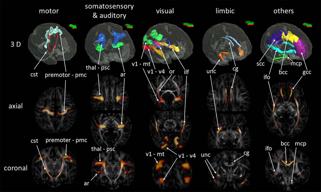
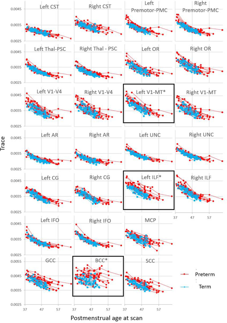
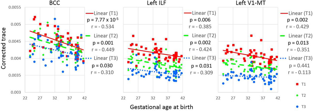
Diffusion tensor imaging (DTI) has been widely used to investigate the development of the neonatal and infant brain, and deviations related to various diseases or medical conditions like preterm birth. In this study, we created a probabilistic map of fiber pathways with known associated functions, on a published neonatal multimodal atlas. The pathways-of-interest include the superficial white matter (SWM) fibers just beneath the specific cytoarchitectonically defined cortical areas, which were difficult to evaluate with existing DTI analysis methods. The Jülich cytoarchitectonic atlas was applied to define cortical areas related to specific brain functions, and the Dynamic Programming (DP) method was applied to delineate the white matter pathways traversing through the SWM. Probabilistic maps were created for pathways related to motor, somatosensory, auditory, visual, and limbic functions, as well as major white matter tracts, such as the corpus callosum, the inferior fronto-occipital fasciculus, and the middle cerebellar peduncle, by delineating these structures in eleven healthy term-born neonates. In order to characterize maturation-related changes in diffusivity measures of these pathways, the probabilistic maps were then applied to DTIs of 49 healthy infants who were longitudinally scanned at three time-points, approximately five weeks apart. First, we investigated the normal developmental pattern based on 19 term-born infants. Next, we analyzed 30 preterm-born infants to identify developmental patterns related to preterm birth. Last, we investigated the difference in diffusion measures between these groups to evaluate the effects of preterm birth on the development of these functional pathways. Term-born and preterm-born infants both demonstrated a time-dependent decrease in diffusivity, indicating postnatal maturation in these pathways, with laterality seen in the corticospinal tract and the optic radiation. The comparison between term- and preterm-born infants indicated higher diffusivity in the preterm-born infants than in the term-born infants in three of these pathways: the body of the corpus callosum; the left inferior longitudinal fasciculus; and the pathway connecting the left primary/secondary visual cortices and the motion-sensitive area in the occipitotemporal visual cortex (V5/MT+). Probabilistic maps provided an opportunity to investigate developmental changes of each white matter pathway. Whether alterations in white matter pathways can predict functional outcomes will be further investigated in a follow-up study.
Keywords: Diffusion tensor imaging; Functional pathway; Neonate; Preterm birth; Probabilistic map; Tractography.
Copyright © 2015 Elsevier Inc. All rights reserved.
Figures
References
-
- Alexandrou G, Martensson G, Skiold B, Blennow M, Aden U, Vollmer B. White matter microstructure is influenced by extremely preterm birth and neonatal respiratory factors. Acta Paediatr. 2014;103:48–56. - PubMed
-
- Amunts K, Malikovic A, Mohlberg H, Schormann T, Zilles K. Brodmann’s areas 17 and 18 brought into stereotaxic space-where and how variable? Neuroimage. 2000;11:66–84. - PubMed
-
- Anjari M, Srinivasan L, Allsop JM, Hajnal JV, Rutherford MA, Edwards AD, Counsell SJ. Diffusion tensor imaging with tract-based spatial statistics reveals local white matter abnormalities in preterm infants. Neuroimage. 2007;35:1021–1027. - PubMed
-
- Ashtari M, Cottone J, Ardekani BA, Cervellione K, Szeszko PR, Wu J, Chen S, Kumra S. Disruption of white matter integrity in the inferior longitudinal fasciculus in adolescents with schizophrenia as revealed by fiber tractography. Arch Gen Psychiatry. 2007;64:1270–1280. - PubMed
-
- Atkinson J, Anker S, Braddick O, Nokes L, Mason A, Braddick F. Visual and visuospatial development in young children with Williams syndrome. Dev Med Child Neurol. 2001;43:330–337. - PubMed
Publication types
MeSH terms
Grants and funding
- G12 MD007601/MD/NIMHD NIH HHS/United States
- G12MD007601-26/MD/NIMHD NIH HHS/United States
- R01HD065955/HD/NICHD NIH HHS/United States
- UL1 TR001079/TR/NCATS NIH HHS/United States
- 1UL1TR001079/TR/NCATS NIH HHS/United States
- K24 DA016170/DA/NIDA NIH HHS/United States
- U54 NS056883/NS/NINDS NIH HHS/United States
- U54NS056883/NS/NINDS NIH HHS/United States
- 2K24DA16170/DA/NIDA NIH HHS/United States
- P41EB015909/EB/NIBIB NIH HHS/United States
- R01 HD065955/HD/NICHD NIH HHS/United States
- P41 EB015909/EB/NIBIB NIH HHS/United States
LinkOut - more resources
Full Text Sources
Other Literature Sources
Medical
