. 2016 Feb 29;44(4):1732-45.
doi: 10.1093/nar/gkv1499. Epub 2015 Dec 27.

Phosphorylation of Ku dictates DNA double-strand break (DSB) repair pathway choice in S phase

Affiliations

Phosphorylation of Ku dictates DNA double-strand break (DSB) repair pathway choice in S phase

Kyung-Jong Lee et al. Nucleic Acids Res. .

Abstract

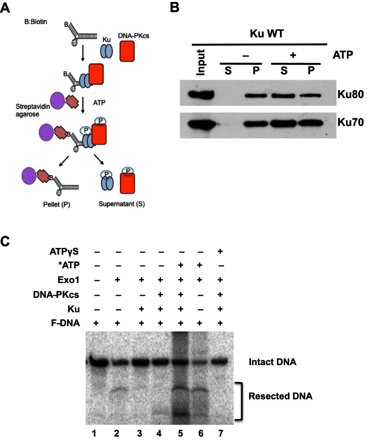
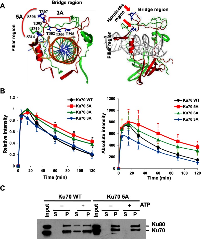
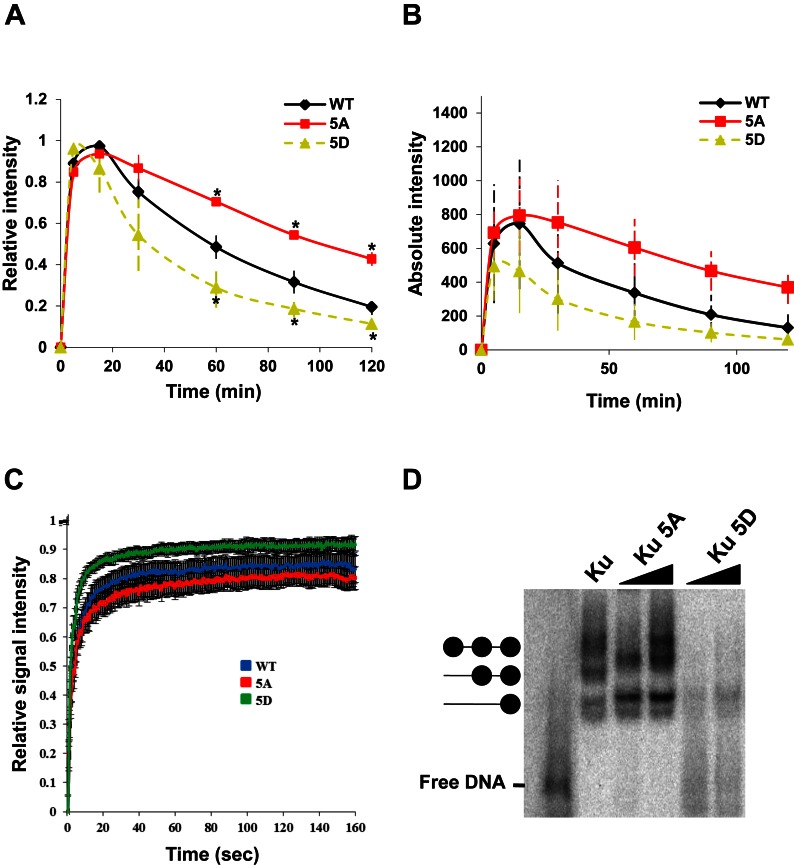
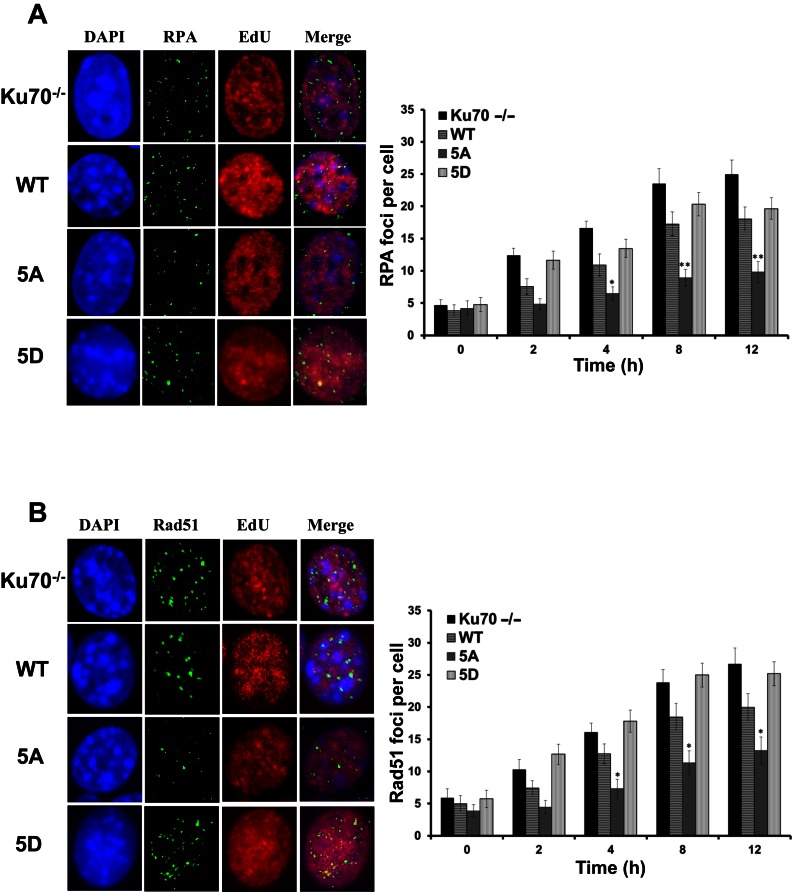
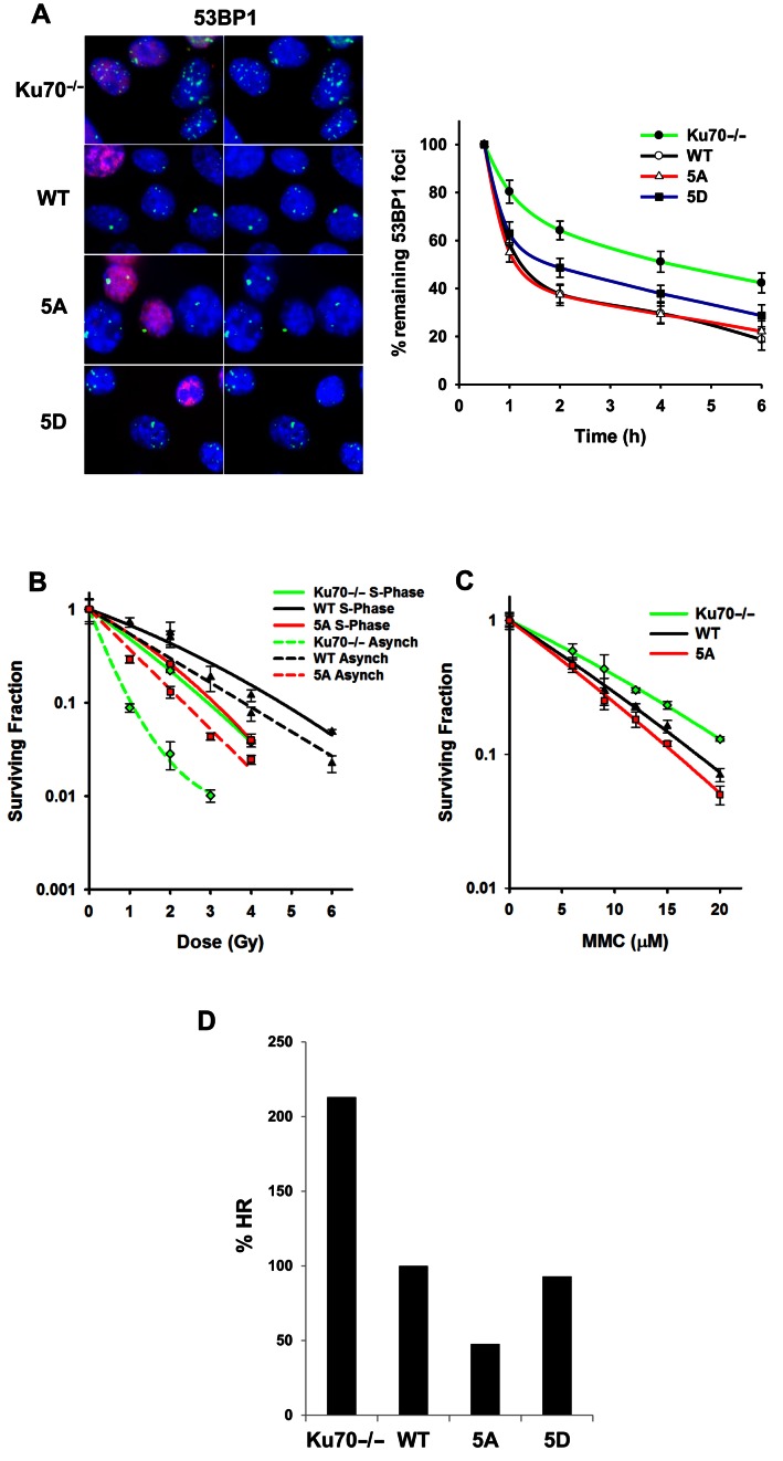
Multiple DNA double-strand break (DSB) repair pathways are active in S phase of the cell cycle; however, DSBs are primarily repaired by homologous recombination (HR) in this cell cycle phase. As the non-homologous end-joining (NHEJ) factor, Ku70/80 (Ku), is quickly recruited to DSBs in S phase, we hypothesized that an orchestrated mechanism modulates pathway choice between HR and NHEJ via displacement of the Ku heterodimer from DSBs to allow HR. Here, we provide evidence that phosphorylation at a cluster of sites in the junction of the pillar and bridge regions of Ku70 mediates the dissociation of Ku from DSBs. Mimicking phosphorylation at these sites reduces Ku's affinity for DSB ends, suggesting that phosphorylation of Ku70 induces a conformational change responsible for the dissociation of the Ku heterodimer from DNA ends. Ablating phosphorylation of Ku70 leads to the sustained retention of Ku at DSBs, resulting in a significant decrease in DNA end resection and HR, specifically in S phase. This decrease in HR is specific as these phosphorylation sites are not required for NHEJ. Our results demonstrate that the phosphorylation-mediated dissociation of Ku70/80 from DSBs frees DNA ends, allowing the initiation of HR in S phase and providing a mechanism of DSB repair pathway choice in mammalian cells.


Figures

Figure 1.
Figure 1.
Ku phosphorylation mediates its dissociation from DNA ends. (A) Schematic diagram of the in vitro DNA-Ku pull-down assay. (B) Phosphorylation of Ku led to its displacement from dsDNA ends in vitro. Purified Ku and DNA-PKcs were incubated with biotin-labeled forked dsDNA followed by the addition of ATP to allow phosphorylation of Ku by DNA-PKcs. The dsDNA was pulled down via streptavidin agarose. Dissociation from the forked dsDNA was assessed with Ku either still bound (pellet, P) or dissociated from the dsDNA (supernatant, S). (C) In vitro DNA resection assay. Exonuclease 1 (Exo1) digests the fork DNA substrate (lane 2 and 6). In the presence of Ku or the DNA-PK complex (DNA-PKcs and Ku), Exo1 could not resect the DNA (lane 3 and 4). The addition of ATP to the forked DNA substrate in the presence of the DNA-PK complex resulted in phosphorylation of Ku, leading to the dissociation of Ku and freeing DNA ends for Exo1-mediated resection (lane 5). Non-hydrolyzable ATP (ATPγS) was used as a control for phosphorylation mediating the freeing of the dsDNA ends (lane 7).
Figure 2.
Figure 2.
Phosphorylation of Ku70 is required for the dissociation of Ku from DSBs. (A) Structure of Ku70/80 with the DNA. Ku70 is indicated in red, while Ku80 is depicted in green. The locations of putative phosphorylation sites in Ku70 are denoted. Ku70 8A cluster sites include both 5A and 3A phosphorylation clusters. (B) Relative and absolute fluorescence intensity of YFP-tagged Ku70 and mutant proteins in Ku70−/− MEFs at DSBs after micro-irradiation. Student's t-test was performed to assess statistical significance (*P < 0.05). (C) Phosphorylation at the 5A sites is required for Ku's dissociation from DSBs. Results were similar to those indicated in 1B, but experiment was performed with purified Ku heterodimer with Ku80 with either wild-type Ku70 or 5A.
Figure 3.
Figure 3.
Mimicking phosphorylation of the five putative sites reduced Ku's DNA binding affinity. (A) Relative and (B) Absolute fluorescence intensity of YFP-tagged Ku70 and mutant proteins in Ku70−/− MEFs at DSBs after micro-irradiation. Student's t-test was performed to assess statistical significance (*P < 0.05). (C) FRAP curves of Ku70 WT, 5A and 5D at the DSB site. Each data point is the average of 15 independent, normalized measurements. Error bars represent the standard deviation (SD). Fluorescence images were captured every 3 s up to 160 s. Pre-bleach intensity levels were normalized to 1, while post-bleach intensity levels were normalized to 0. Zero second represents the time of bleaching signal. (D) Electrophoretic mobility shift assays with purified wild-type Ku80 and wild-type Ku70, 5A or 5D with forked dsDNA.
Figure 4.
Figure 4.
Blocking phosphorylation of Ku70 attenuates initiation of end resection and onset of homologous recombination. Panel of images depict (A) RPA and (B) Rad51 foci in EdU positive cells after 8 h after 8Gy gamma-ray exposure, respectively (Left panels). Immunostaining of Ku70−/− MEFs or Ku70−/− MEFs complemented with Ku70 wild-type, 5A or 5D after 8Gy of γ-rays. Cells were pre-extracted and fixed 2, 4, 8, or 12 h after IR and immunostained for RPA or Rad51. RPA and Rad51 foci were counted for each cell and averaged (Right panels). Student's t-test was performed to assess statistical significance (*P < 0.01 and **P < 0.001). Error bars denote standard error of the mean (SEM) for at least 3 independent experiments.
Figure 5.
Figure 5.
Ablation of Ku70 phosphorylation results in attenuation of homologous recombination. (A) Images depicting 53BP1 foci, enumerated in EdU negative G1 phase cells after 6 h of 1Gy gamma-ray exposure (Left panel). Ku70−/− MEFs or Ku70−/− MEFs complemented with Ku70 wild-type, 5A or 5D were irradiated with 1Gy and 53BP1. Foci formation was assessed 1, 2, 4, and 6 h later (Right panel). Data were normalized to 53BP1 foci number enumerated at 30 min post IR. Remaining foci number per time point assessed was calculated and plotted. Error bars denote SEM of at least three independent experiments. (B) Colony formation assays were performed to compare radiation sensitivities of Ku70−/− MEFs or Ku70−/− MEFs complemented with Ku70 wild-type or 5A in S phase or as an asynchronous cell population. Cell lines were left cycling or were synchronized by the double thymidine block method and then released. Subsequently, cells were irradiated at the indicated doses and plated for analysis of survival and colony-forming ability. Error bars denote SD values. (C) Colony formation assays were performed to compare mitomycin C (MMC) sensitivity of Ku70−/− MEFs or Ku70−/− MEFs complemented with Ku70 wild-type or 5A. Cell lines were treated with the indicated concentrations of MMC and plated for analysis of survival and colony-forming ability. Error bars denote SD values. (D) Ku70−/− MEFs, containing one copy of the HR reporter assay vector integrated into the genome, were either uncomplemented or complemented with FLAG-tagged Ku70 wild-type, 5A or 5D. I-SceI was transiently expressed and induced a DSB at the I-SceI restriction site. Cells were analyzed by flow cytometry and the percent HR value was calculated as GFP / (RFP + GFP). The percent HR value of wild-type was normalized to 100% and the percent HR of Ku70−/− MEFs, 5A and 5D cells were calculated and plotted.

References

    1. Jackson S.P., Bartek J. The DNA-damage response in human biology and disease. Nature. 2009;461:1071–1078. - PMC - PubMed
    1. Schipler A., Iliakis G. DNA double-strand-break complexity levels and their possible contributions to the probability for error-prone processing and repair pathway choice. Nucleic Acids Res. 2013;41:7589–7605. - PMC - PubMed
    1. Hoeijmakers J.H. Genome maintenance mechanisms for preventing cancer. Nature. 2001;411:366–374. - PubMed
    1. Goodarzi A.A., Jeggo P.A. The repair and signaling responses to DNA double-strand breaks. Adv. Genet. 2013;82:1–45. - PubMed
    1. Thompson L.H. Recognition, signaling, and repair of DNA double-strand breaks produced by ionizing radiation in mammalian cells: the molecular choreography. Mutat. Res. 2012;751:158–246. - PubMed

Publication types

LinkOut - more resources