

A Herbal Formula, Atofreellage, Ameliorates Atopic Dermatitis-Like Skin Lesions in an NC/Nga Mouse Model
- PMID: 26712731
- PMCID: PMC6273903
- DOI: 10.3390/molecules21010035
A Herbal Formula, Atofreellage, Ameliorates Atopic Dermatitis-Like Skin Lesions in an NC/Nga Mouse Model
Abstract
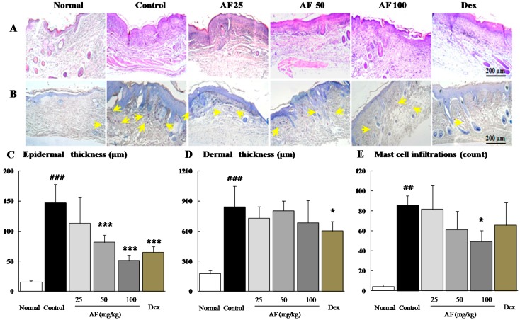
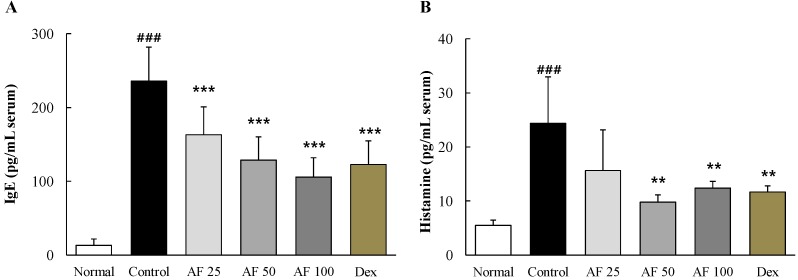
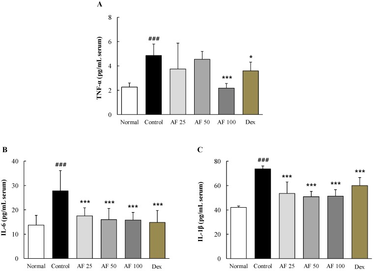
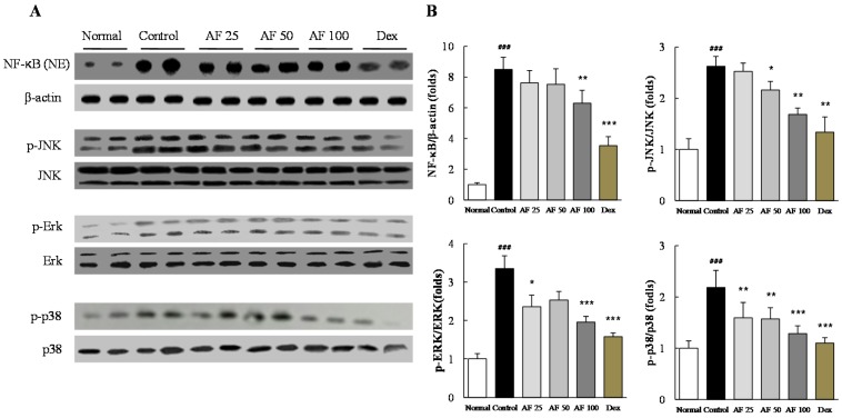
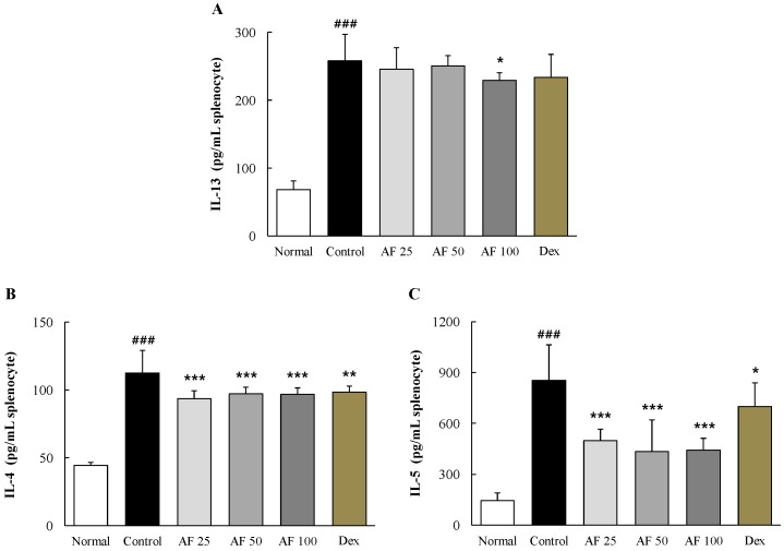
We evaluated the anti-atopic dermatitis (AD) effect of Atofreellage (AF), a herbal formula composed of 10 medicinal plants. AD was induced on the dorsal skin areas of NC/Nga mice (male, seven weeks old) by daily application of 2,4-dinitrochlorobenzene (DNCB) for five weeks. After three weeks of DNCB application, 200 μL of AF (0, 25, 50 or 100 mg/mL) was applied to the skin lesions. Histological findings, blood cell populations, serum levels of immunoglobulin E (IgE), histamine, pro-inflammatory cytokines, and inflammatory signaling in the skin tissue, and T-helper cell type 2 (Th₂)-related cytokines in splenocytes were analyzed. Histopathological findings showed AF treatment notably attenuated the thickness of dorsal skin, and eosinophil infiltration. AF treatment (especially 100 mg/mL) also demonstrably ameliorated the blood cell population abnormalities, as the notable elevation of serum concentrations of IgE, histamine, TNF-α, IL-6 and IL-1β were remarkably normalized by AF treatment. Western blot analysis evidenced the apparent normalization of inflammatory signals (ERK, p38 MAP kinase, JNK, and NF-κB) in the skin tissue. Additionally, AF treatment notably attenuated the activation of Th₂-dominant cytokines (IL-13, IL-4, and IL-5) in Con A-treated splenocytes in an ex vivo assay. In conclusion, this study provides experimental evidence for the clinical relevance of Atofreellage.
Keywords: NC/Nga murine model; atopic dermatitis; herbal medicine; inflammation.
Conflict of interest statement
The authors declare no conflict of interest.
Figures
Similar articles
-
Pyrus ussuriensis Maxim. leaves extract ameliorates DNCB-induced atopic dermatitis-like symptoms in NC/Nga mice.Phytomedicine. 2018 Sep 15;48:76-83. doi: 10.1016/j.phymed.2018.05.006. Epub 2018 May 8. Phytomedicine. 2018. PMID: 30195883
-
Inhibitory effects of Solanum tuberosum L. var. vitelotte extract on 2,4-dinitrochlorobenzene-induced atopic dermatitis in mice.J Pharm Pharmacol. 2014 Sep;66(9):1303-16. doi: 10.1111/jphp.12254. Epub 2014 Jun 24. J Pharm Pharmacol. 2014. PMID: 24961480
-
Qing-Re-Chu-shi decoction ameliorates 2,4-dinitrochlorobenzene-induced atopic dermatitis in NC/Nga mice through anti-inflammation and immunoregulatory mechanisms.J Ethnopharmacol. 2024 Apr 6;323:117702. doi: 10.1016/j.jep.2024.117702. Epub 2024 Jan 3. J Ethnopharmacol. 2024. PMID: 38176665
-
[Atopic dermatitis and cathepsin E].Nihon Yakurigaku Zasshi. 2003 Jul;122(1):15-20. doi: 10.1254/fpj.122.15. Nihon Yakurigaku Zasshi. 2003. PMID: 12843568 Review. Japanese.
-
Herbal Medicines Prevent the Development of Atopic Dermatitis by Multiple Mechanisms.Chin J Integr Med. 2019 Feb;25(2):151-160. doi: 10.1007/s11655-015-2438-1. Epub 2016 Jan 6. Chin J Integr Med. 2019. PMID: 26740223 Review.
Cited by
-
Integrated Omics Analysis Uncovers the Culprit behind Exacerbated Atopic Dermatitis in a Diet-Induced Obesity Model.Int J Mol Sci. 2024 Apr 9;25(8):4143. doi: 10.3390/ijms25084143. Int J Mol Sci. 2024. PMID: 38673730 Free PMC article.
-
Management of Atopic Dermatitis Via Oral and Topical Administration of Herbs in Murine Model: A Systematic Review.Front Pharmacol. 2022 May 24;13:785782. doi: 10.3389/fphar.2022.785782. eCollection 2022. Front Pharmacol. 2022. PMID: 35685636 Free PMC article.
References
-
- Berke R., Singh A., Guralnick M. Atopic dermatitis: An overview. Am. Fam. Phys. 2012;86:35–42. - PubMed
Publication types
MeSH terms
Substances
LinkOut - more resources
Full Text Sources
Other Literature Sources
Research Materials
Miscellaneous
