

Novel Insights into Guide RNA 5'-Nucleoside/Tide Binding by Human Argonaute 2
- PMID: 26712743
- PMCID: PMC4730269
- DOI: 10.3390/ijms17010022
Novel Insights into Guide RNA 5'-Nucleoside/Tide Binding by Human Argonaute 2
Abstract
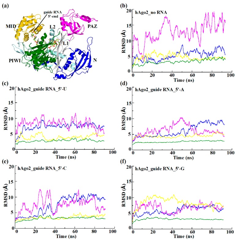
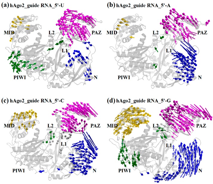
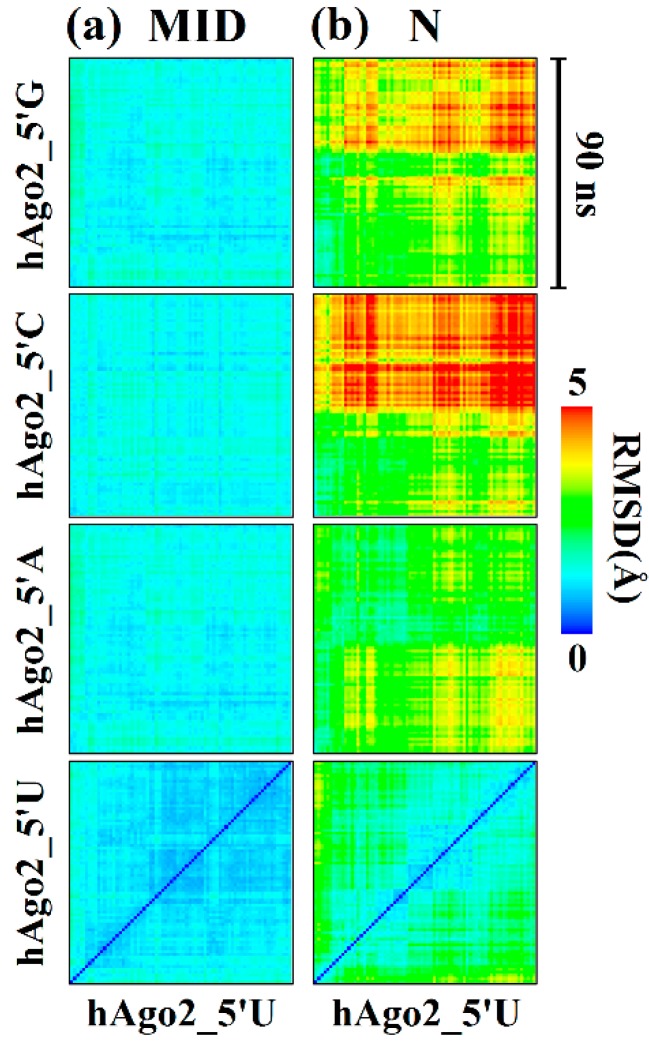
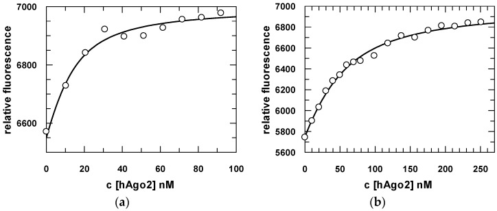
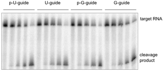
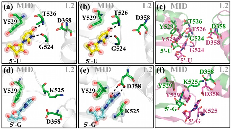
The human Argonaute 2 (hAgo2) protein is a key player of RNA interference (RNAi). Upon complex formation with small non-coding RNAs, the protein initially interacts with the 5'-end of a given guide RNA through multiple interactions within the MID domain. This interaction has been reported to show a strong bias for U and A over C and G at the 5'-position. Performing molecular dynamics simulations of binary hAgo2/OH-guide-RNA complexes, we show that hAgo2 is a highly flexible protein capable of binding to guide strands with all four possible 5'-bases. Especially, in the case of C and G this is associated with rather large individual conformational rearrangements affecting the MID, PAZ and even the N-terminal domains to different degrees. Moreover, a 5'-G induces domain motions in the protein, which trigger a previously unreported interaction between the 5'-base and the L2 linker domain. Combining our in silico analyses with biochemical studies of recombinant hAgo2, we find that, contrary to previous observations, hAgo2 is capable of functionally accommodating guide strands regardless of the 5'-base.
Keywords: MD; RNAi; enzyme kinetics; fluorescence spectroscopy; pre-steady-state kinetics.
Figures
Similar articles
-
Exploring the RNA-bound and RNA-free human Argonaute-2 by molecular dynamics simulation method.Chem Biol Drug Des. 2017 Nov;90(5):753-763. doi: 10.1111/cbdd.12997. Epub 2017 May 12. Chem Biol Drug Des. 2017. PMID: 28388001
-
Probing the Binding Interactions between Chemically Modified siRNAs and Human Argonaute 2 Using Microsecond Molecular Dynamics Simulations.J Chem Inf Model. 2017 Apr 24;57(4):883-896. doi: 10.1021/acs.jcim.6b00773. Epub 2017 Mar 27. J Chem Inf Model. 2017. PMID: 28287733
-
Single-molecule FRET uncovers hidden conformations and dynamics of human Argonaute 2.Nat Commun. 2022 Jul 2;13(1):3825. doi: 10.1038/s41467-022-31480-4. Nat Commun. 2022. PMID: 35780145 Free PMC article.
-
Understanding the core of RNA interference: The dynamic aspects of Argonaute-mediated processes.Prog Biophys Mol Biol. 2017 Sep;128:39-46. doi: 10.1016/j.pbiomolbio.2016.09.008. Epub 2016 Sep 30. Prog Biophys Mol Biol. 2017. PMID: 27697475 Review.
-
Conformational Dynamics of Ago-Mediated Silencing Processes.Int J Mol Sci. 2015 Jul 1;16(7):14769-85. doi: 10.3390/ijms160714769. Int J Mol Sci. 2015. PMID: 26140373 Free PMC article. Review.
Cited by
-
RNA-protein complexes and force field polarizability.Front Chem. 2023 Jun 22;11:1217506. doi: 10.3389/fchem.2023.1217506. eCollection 2023. Front Chem. 2023. PMID: 37426330 Free PMC article.
-
Autonomous Generation and Loading of DNA Guides by Bacterial Argonaute.Mol Cell. 2017 Mar 16;65(6):985-998.e6. doi: 10.1016/j.molcel.2017.01.033. Epub 2017 Mar 2. Mol Cell. 2017. PMID: 28262506 Free PMC article.
-
RNA-induced initiation of transcriptional silencing (RITS) complex structure and function.RNA Biol. 2019 Sep;16(9):1133-1146. doi: 10.1080/15476286.2019.1621624. Epub 2019 Jun 18. RNA Biol. 2019. PMID: 31213126 Free PMC article. Review.
-
Comparative structural insights and functional analysis for the distinct unbound states of Human AGO proteins.Sci Rep. 2025 Mar 19;15(1):9432. doi: 10.1038/s41598-025-91849-5. Sci Rep. 2025. PMID: 40108192 Free PMC article.
-
Exploring PAZ/3'-overhang interaction to improve siRNA specificity. A combined experimental and modeling study.Chem Sci. 2018 Jan 15;9(8):2074-2086. doi: 10.1039/c8sc00010g. eCollection 2018 Feb 28. Chem Sci. 2018. PMID: 29719684 Free PMC article.
References
Publication types
MeSH terms
Substances
LinkOut - more resources
Full Text Sources
Other Literature Sources
