Review
. 2015 Dec 25;5(1):1.
doi: 10.3390/jcm5010001.

Epithelial-Mesenchymal Transitions during Neural Crest and Somite Development

Affiliations
Review

Epithelial-Mesenchymal Transitions during Neural Crest and Somite Development

Chaya Kalcheim. J Clin Med. .

Abstract

Epithelial-to-mesenchymal transition (EMT) is a central process during embryonic development that affects selected progenitor cells of all three germ layers. In addition to driving the onset of cellular migrations and subsequent tissue morphogenesis, the dynamic conversions of epithelium into mesenchyme and vice-versa are intimately associated with the segregation of homogeneous precursors into distinct fates. The neural crest and somites, progenitors of the peripheral nervous system and of skeletal tissues, respectively, beautifully illustrate the significance of EMT to the above processes. Ongoing studies progressively elucidate the gene networks underlying EMT in each system, highlighting the similarities and differences between them. Knowledge of the mechanistic logic of this normal ontogenetic process should provide important insights to the understanding of pathological conditions such as cancer metastasis, which shares some common molecular themes.

Keywords: BMP; FGF; N-cadherin; Wnt; cell fate; cell migration; dermis; dermomyotome; neural tube; sclerotome.


Figures

Figure 1
Figure 1
Epithelial to mesenchymal transition (EMT) of neural crest (NC) progenitors: Regulation and cellular dynamics. (A) The medial lip of the nascent dermomyotome (DM) controls the timing of NC delamination. In the early dorsal NT, prior to the onset of NC emigration, levels of Noggin are high thereby inhibiting the activity of BMP4 and NC delamination. The forming medial lip of the DM acts upon the dorsal NT via FGF8 and retinoic acid signaling to inhibit local noggin transcription thus relieving BMP4 which stimulates NC emigration. Scl, sclerotome; (B) Two possible models representing the dynamic dorsalward relocation of NC cells prior to emigration in association with fate restriction. In both models, the emigrating cells are largely fate-restricted. In the model on the left (spatial mechanism), a pattern reflecting the different fates is already apparent in the dorsal NT (color coding). In the model depicted on the right, fate restriction is assigned to a cell upon relocation to the dorsal midline area by a time-dependent mechanism. Abbreviations: DRG, dorsal root ganglia; M, melanocytes; RP, roof plate; SG, sympathetic ganglia; VR, Schwann cells along ventral root.
Figure 2
Figure 2
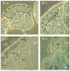
Successive stages in dermomyotome development at flank levels of the axis. Phase contrast images that illustrate: (A) T0, epithelial somite stage with the prospective dermomyotome (DM) highlighted by dashed lines; (B) T1, initial formation of the DM following mesenchymalization of the sclerotome (Scl). At this stage the pioneer (P) myoblasts bend underneath the nascent DM; (C) T2, the DM of a fully dissociated somite in which the primary myotome (M) is well differentiated. Note the well defined medial and lateral edges (DML and VLL, respectively); (D) T3, the DM dissociates into dermis except for the DML and VLL which still remain epithelial (demarcated by dashed lines). See text for precise stages. Abbreviations, DRG, dorsal root ganglion, NT, neural tube. Bar = (A) 8 μM; (B) 15 μM; (C) 22 μM; and (D) 80 μM.

References

    1. Savagner P. Leaving the neighborhood: Molecular mechanisms involved during epithelial-mesenchymal transition. BioEssays. 2004;23:912–923. doi: 10.1002/bies.1132. - DOI - PubMed
    1. Le Douarin N.M., Kalcheim C. The Neural Crest. 2nd ed. Cambridge University Press; New York, NY, USA: 1999.
    1. Bronner-Fraser M. Segregation of cell lineage in the neural crest. Curr. Opin. Genet. Dev. 1993;3:641–647. doi: 10.1016/0959-437X(93)90101-T. - DOI - PubMed
    1. Bronner M.E. Formation and migration of neural crest cells in the vertebrate embryo. Histochem. Cell Biol. 2012;138:179–186. doi: 10.1007/s00418-012-0999-z. - DOI - PMC - PubMed
    1. Weston J.A. The migration and differentiation of neural crest cells. Adv. Morphog. 1970;8:41–114. - PubMed

LinkOut - more resources