. 2015 Dec 29:21:4081-9.
doi: 10.12659/msm.895547.

Niacin Suppresses Progression of Atherosclerosis by Inhibiting Vascular Inflammation and Apoptosis of Vascular Smooth Muscle Cells

Affiliations

Niacin Suppresses Progression of Atherosclerosis by Inhibiting Vascular Inflammation and Apoptosis of Vascular Smooth Muscle Cells

Gang Su et al. Med Sci Monit. .

Abstract

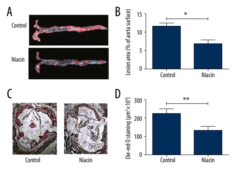
BACKGROUND Niacin is a broad-spectrum lipid-regulating drug used for the clinical therapy of atherosclerosis; however, the mechanisms by which niacin ameliorates atherosclerosis are not clear. MATERIAL AND METHODS The effect of niacin on atherosclerosis was assessed by detection of atherosclerotic lesion area. Adhesion molecules in arterial endothelial cells were determined by using qRT-PCR and Western blot analysis. The levels of serum inflammatory cytokines in ApoE-/- mice were detected by using ELISA. We detected the expression levels of phosphorylated nuclear factors-kB (NF-κB) p65 in aortic endothelial cells of mice using Western blot analysis. Furthermore, we investigated the anti-inflammation effect and endothelium-protecting function of niacin and their regulatory mechanisms in vitro. RESULTS Niacin inhibited the progress of atherosclerosis and decreased the levels of serum inflammatory cytokines and adhesion molecules in ApoE-/- mice. Niacin suppressed the activity of NF-κB and apoptosis of vascular smooth muscle cells (VSMCs). Furthermore, niacin induced phosphorylated focal adhesion kinase (FAK) and FAK inhibitor PF-573228 reduced the level of Bcl-2 and elevated the level of cleaved caspase-3 in VSMCs. CONCLUSIONS Niacin inhibits vascular inflammation and apoptosis of VSMCs via inhibiting the NF-κB signaling and the FAK signaling pathway, respectively, thus protecting ApoE-/- mice against atherosclerosis.


Figures

Figure 1
Figure 1
Niacin inhibits the progression of atherosclerosis in ApoE−/− mice. Representative oil red O-stained aortic arches (A) and cryosections of aortic roots (C) from untreated and niacin-treated ApoE−/− mice. The areas of atherosclerotic plaque were measured by image analysis. Data are presented as average lesion size in percentage of the total surface of the aorta (B) or as the absolute lesion area on the aortic root (D). Data are presented as mean ±SD (n=6).* P<0.05, ** P<0.01.
Figure 2
Figure 2
Niacin suppresses expression of adhesion molecules in ApoE−/− mice. qRT-PCR and Western blot analysis were used to analyze the mRNA and protein expression levels of adhesion molecules in different treatment groups of mice, respectively. The mRNA (A) and protein (B) level of ICAM-1, E-selectin and VCAM-1 in the mice vascular endothelial cells were obviously decreased after feeding niacin. Relative protein levels of ICAM-1, E-selectin and VCAM-1 were quantified using Image-Pro Plus 6.0 software and normalized to GAPDH. Data were expressed as mean ±SD from independent mice (n=6), ** P<0.01.
Figure 3
Figure 3
(A–D) Niacin decreases the levels of serum inflammatory cytokines in mouse models. The levels of IL-1β, IL-6, TNF-α, and MCP-1 in serum of ApoE−/− mice were determined by ELISA after treatment for 10 weeks. Compared with the control group, the levels of serum inflammatory cytokines were significantly lower in niacin group. Data presented as mean ±SD (n=6). * P<0.05; ** P<0.01.
Figure 4
Figure 4
The molecular mechanism of niacin on NF-κB signaling pathway. (A) Western blot analysis was performed to detect the protein expression levels in aortic endothelial cells of ApoE−/− mice. Niacin significantly decreased the phosphorylation of p65. (B, C) Niacin reduced the expression levels of TNF-α, IL-1β, IL-6 and MCP-1. (D, E) Niacin suppressed the level of p-p65. (F) Luciferase reporter assay were used to detect the activity of NF-κB in HAECs. The relative luciferase activity was increased after treatment with TNF-α. However, niacin decreased the relative luciferase activity, showing that niacin inhibits the NF-κB signaling pathway. Data presented as mean ±SD (n=4). * P<0.05, ** P<0.01, *** P<0.001.
Figure 5
Figure 5
Niacin inhibits oxLDL-induced apoptosis of VSMCs through knocking the FAK signaling pathway. (A) MDA level increased in cells exposed to oxLDL. (B) The apoptosis rate was increased after treatment with oxLDL, but niacin resisted oxLDL-induced apoptosis of VSMCs. (B) Niacin reduced the apoptosis rate of VSMCs induced by oxLDL. (C) Niacin promoted the expression of Bcl-2 but repressed the expression of cleaved caspase-3. (D) Niacin induced the level of p-FAK in a concentration-dependent manner. (E) VSMCs were treated with 50 μM FAK inhibitor PF-573228 for 24 h. Blocking the FAK signaling decreased the expression level of Bcl-2 but increased the expression level of cleaved caspase-3. Data presented as mean ±SD (n=4). * P<0.05, ** P<0.01, *** P<0.001.

References

    1. Libby P, Ridker PM, Maseri A. Inflammation and atherosclerosis. Circulation. 2002;105:1135–43. - PubMed
    1. Libby P. Inflammation in atherosclerosis. Arterioscl Throm Vas. 2012;32:2045–51. - PMC - PubMed
    1. Li D, Yang B, Mehta JL. Ox-LDL induces apoptosis in human coronary artery endothelial cells: role of PKC PTK bcl-2 and Fas. Am J Physiol Heart Circ Physiol. 1998;275:H568–76. - PubMed
    1. Chen J, Mehta JL, Haider N, et al. Role of caspases in Ox-LDL – induced apoptotic cascade in human coronary artery endothelial cells. Circ Res. 2004;94:370–76. - PubMed
    1. Skeoch S, Haque S, Pemberton P, Bruce I. Cell adhesion molecules as potential biomarkers of nephritis damage and accelerated atherosclerosis in patients with SLE. Lupus. 2014;23:819–24. - PMC - PubMed

Publication types

MeSH terms