Review
. 2016 Jan;147(1):1-24.
doi: 10.1085/jgp.201511492.

The hitchhiker's guide to the voltage-gated sodium channel galaxy

Affiliations
Review

The hitchhiker's guide to the voltage-gated sodium channel galaxy

Christopher A Ahern et al. J Gen Physiol. 2016 Jan.

Abstract

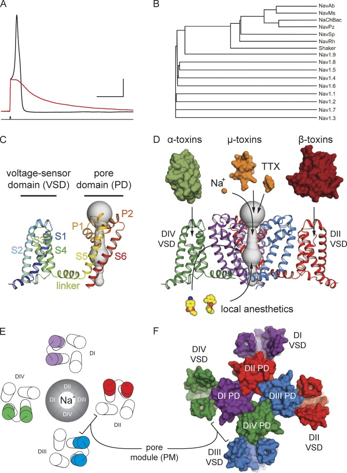
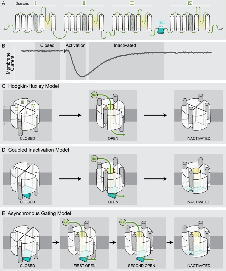
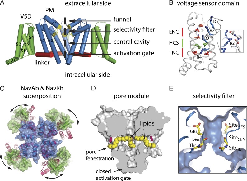
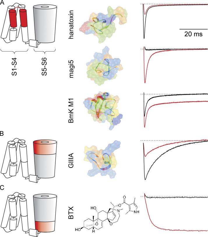
Eukaryotic voltage-gated sodium (Nav) channels contribute to the rising phase of action potentials and served as an early muse for biophysicists laying the foundation for our current understanding of electrical signaling. Given their central role in electrical excitability, it is not surprising that (a) inherited mutations in genes encoding for Nav channels and their accessory subunits have been linked to excitability disorders in brain, muscle, and heart; and (b) Nav channels are targeted by various drugs and naturally occurring toxins. Although the overall architecture and behavior of these channels are likely to be similar to the more well-studied voltage-gated potassium channels, eukaryotic Nav channels lack structural and functional symmetry, a notable difference that has implications for gating and selectivity. Activation of voltage-sensing modules of the first three domains in Nav channels is sufficient to open the channel pore, whereas movement of the domain IV voltage sensor is correlated with inactivation. Also, structure-function studies of eukaryotic Nav channels show that a set of amino acids in the selectivity filter, referred to as DEKA locus, is essential for Na(+) selectivity. Structures of prokaryotic Nav channels have also shed new light on mechanisms of drug block. These structures exhibit lateral fenestrations that are large enough to allow drugs or lipophilic molecules to gain access into the inner vestibule, suggesting that this might be the passage for drug entry into a closed channel. In this Review, we will synthesize our current understanding of Nav channel gating mechanisms, ion selectivity and permeation, and modulation by therapeutics and toxins in light of the new structures of the prokaryotic Nav channels that, for the time being, serve as structural models of their eukaryotic counterparts.


Figures

Figure 1.
Figure 1.
Nav channel function, family tree, and structural architecture. (A) Evoked action potential recorded from a mouse DRG neuron at room temperature before (black) and after (red) the application of 1 µM TTX. X axis is 30 ms, and y axis is 20 mV. (B) A phylogenetic tree of Nav channels as well as Shaker obtained using Vector NTI AlignX software. (C) The side view of a signal subunit of the NavAb channel homotetramer (Protein Data Bank accession no. 3RVY) in ribbon style is colored from N terminus (blue) to C terminus (red). This view highlights the VSD as a modular four-helix bundle. (D) Side view of the NavAb channel with the front VSD and pore domain removed for clarity. For illustrative purposes, NavAb is colored according to a pseudotetrameric arrangement expected for eukaryotic Nav cannels. Representative classes of protein toxins (α, β, and µ), small molecule toxins (TTX), as well select small molecule drugs (lidocaine and benzocaine) are represented with arrows pointing to their presumed canonical binding sites on the channel. (E) Top-view schematic of a eukaryotic Nav channel with the S3b–S4 region of the VSDs from different domains is highlighted in different colors. The ion-conducting Na+ pore is found in the center of this view. (F) A structural top view of the NavAb channel colored according to a pseudotetrameric arrangement expected for a eukaryotic Nav channel (as in D). This subunit coloring highlights the “domain-swapped arrangement” of the VSDs around the PM observed for all voltage-gated ion channels.
Figure 2.
Figure 2.
Schematic representation of gating models of eukaryotic sodium channels. (A) Transmembrane topology of a eukaryotic Nav channel. The S4 voltage-sensing segment is shaded in gray, and the P-loop constitutes the selectivity filter region. The inactivation motif (cerulean-colored box) is the loop connecting domains III and IV. (B) Representative membrane currents through a voltage-activated sodium channel in response to a depolarizing pulse from a holding potential of −90 mV. The start of the depolarization pulse is represented as a break, and the gating current component has been subtracted. (C) Schematic rendering of the original HH model of sodium channel gating. Rapid activation of three “m” particles is sufficient for the channel to open, and slower activation of the “h” particle causes the channel to inactivate. (D) In the coupled inactivation model, activation of all four voltage sensors contributes to the channel opening. Inactivation results from binding of the inactivation lid to its receptor in the pore, which becomes accessible in the open state. (E) According to the asynchronous gating model, the activation of the first three VSDs of the sodium channel is sufficient to open the channel. Slow activation of the domain IV voltage sensor results in a secondary open state and makes the receptor for inactivation lid accessible.
Figure 3.
Figure 3.
Overview of BacNav crystal structures. (A) Side view of the NavAb channel (Protein Data Bank accession no. 3RVY) with the VSDs colored green, the S4–S5 linkers colored red, and the PM colored blue. The selectivity filter motif in all four subunits is colored yellow. Main regions within the pore structure are indicated, and the front VSD and pore domain are removed for clarity. (B) Voltage-sensor domain from NavAb highlights conserved structural and functional features within the VSD including the hydrophobic constriction site (HCS) and the intracellular and extracellular negative charge clusters (INC and ENC). The gating charges (arginine residues, R1–R4) are shown in blue sticks. (Inset) The R2 arginine gating charge hydrogen bonding with a backbone carbonyl from the S3 helix is highlighted. (C) Superposition of the NavAb and NavRh (Protein Data Bank accession no. 4DXW) channel pores (colored blue and pink, respectively) indicates the possibility of a significant movement of the VSDs within the plane of the membrane. (D) Side-view section of the NavAb channel shows locations of bound phospholipids (yellow spheres) within the PM and their penetration through the pore fenestrations. The side chain of Phe203 is shown in pink stick representation for reference, and the closed intracellular activation gate formed by the S6 helices is indicated. (E) Side view sectioned through the PM of NavAb shows three coordination sites identified within BacNav selectivity filters. From the extracellular to intracellular side, these sites are: SiteHFS, SiteCEN, and SiteIN. The approximate positions of the Thr (T), Leu (L), and Glu (E) backbone or side-chain atoms from the conserved TLESW selectivity motif are also indicated.
Figure 4.
Figure 4.
Interactions between animal toxins and Nav channels. (A; left) Side view of a Nav channel cartoon indicating the paddle motif (indicated in red) as the binding site for hanatoxin from the Grammostola rosea tarantula, Magi5 from the Hexathelidae spider Macrothele gigas, and BmK M1 from the Buthus martensii Karsch scorpion. (Middle) Structures of the three toxins colored according to residue class (green, hydrophobic; blue, positively charged; red, negatively charged; orange, polar). Toxin backbone is also shown. (Right) Effect of 100 nM hanatoxin (channel opening is inhibited), 1 µM Magi5 (channel opens at voltages where it is normally closed), and 100 nM BmK M1 (channel fast inactivation is inhibited) on rNav1.2a channels expressed in Xenopus laevis oocytes and recorded with the two-electrode voltage-clamp technique. Despite binding to a similar region on the Nav channel, each toxin has a different effect on channel opening or closing. Black trace represents control conditions, and red trace represents toxin. (B) Effect of 30 nM cone snail toxin GIIIA on rNav1.4-mediated currents recorded from Xenopus laevis oocytes. GIIIA blocks Na+ flow by binding to the outer region of the pore mouth. (C) Effect of 1 µM BTX, isolated from the poison dart frog, on rNav1.8 channels expressed in Xenopus laevis oocytes. BTX binds to the inner region of the pore to drastically modify Nav channel gating. Shown is the ability of BTX to open Nav channels at voltages where they are normally closed and to inhibit fast inactivation. Black trace represents control conditions, and red trace represents toxin.

References

    1. Abriel H., and Kass R.S.. 2005. Regulation of the voltage-gated cardiac sodium channel Nav1.5 by interacting proteins. Trends Cardiovasc. Med. 15:35–40. 10.1016/j.tcm.2005.01.001 - DOI - PubMed
    1. Agnew W.S., Levinson S.R., Brabson J.S., and Raftery M.A.. 1978. Purification of the tetrodotoxin-binding component associated with the voltage-sensitive sodium channel from Electrophorus electricus electroplax membranes. Proc. Natl. Acad. Sci. USA. 75:2606–2610. 10.1073/pnas.75.6.2606 - DOI - PMC - PubMed
    1. Ahern C.A., Eastwood A.L., Dougherty D.A., and Horn R.. 2008. Electrostatic contributions of aromatic residues in the local anesthetic receptor of voltage-gated sodium channels. Circ. Res. 102:86–94. 10.1161/CIRCRESAHA.107.160663 - DOI - PubMed
    1. Ahuja S., Mukund S., Deng L., Khakh K., Chang E., Ho H., Shriver S., Young C., Lin S., Johnson J.P. Jr., et al. 2015. Structural basis of Nav1.7 inhibition by an isoform-selective small molecule antagonist. Science. 350:1491. - PubMed
    1. Aldrich R.W., and Stevens C.F.. 1983. Inactivation of open and closed sodium channels determined separately. Cold Spring Harb. Symp. Quant. Biol. 48:147–153. 10.1101/SQB.1983.048.01.017 - DOI - PubMed

Publication types