. 2015 Dec 28;198(5):808-15.
doi: 10.1128/JB.00849-15.

Permuting the PGF Signature Motif Blocks both Archaeosortase-Dependent C-Terminal Cleavage and Prenyl Lipid Attachment for the Haloferax volcanii S-Layer Glycoprotein

Affiliations

Permuting the PGF Signature Motif Blocks both Archaeosortase-Dependent C-Terminal Cleavage and Prenyl Lipid Attachment for the Haloferax volcanii S-Layer Glycoprotein

Mohd Farid Abdul Halim et al. J Bacteriol. .

Abstract

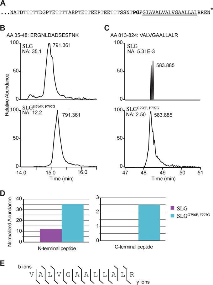
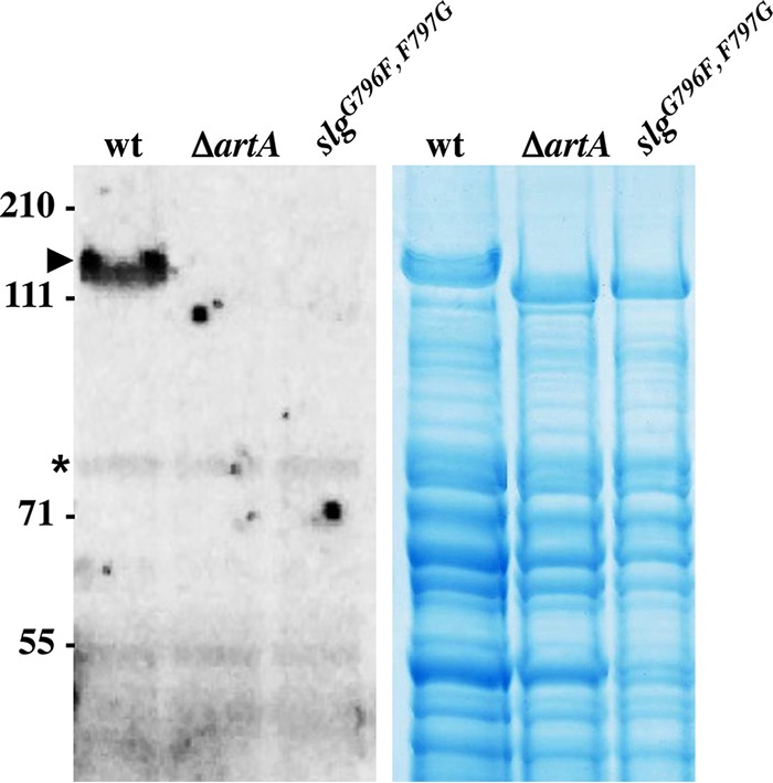
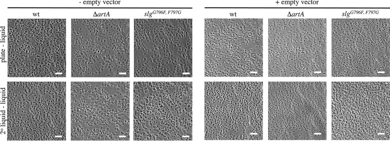
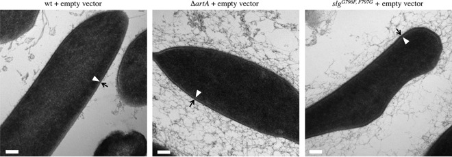
For years, the S-layer glycoprotein (SLG), the sole component of many archaeal cell walls, was thought to be anchored to the cell surface by a C-terminal transmembrane segment. Recently, however, we demonstrated that the Haloferax volcanii SLG C terminus is removed by an archaeosortase (ArtA), a novel peptidase. SLG, which was previously shown to be lipid modified, contains a C-terminal tripartite structure, including a highly conserved proline-glycine-phenylalanine (PGF) motif. Here, we demonstrate that ArtA does not process an SLG variant where the PGF motif is replaced with a PFG motif (slg(G796F,F797G)). Furthermore, using radiolabeling, we show that SLG lipid modification requires the PGF motif and is ArtA dependent, lending confirmation to the use of a novel C-terminal lipid-mediated protein-anchoring mechanism by prokaryotes. Similar to the case for the ΔartA strain, the growth, cellular morphology, and cell wall of the slg(G796F,F797G) strain, in which modifications of additional H. volcanii ArtA substrates should not be altered, are adversely affected, demonstrating the importance of these posttranslational SLG modifications. Our data suggest that ArtA is either directly or indirectly involved in a novel proteolysis-coupled, covalent lipid-mediated anchoring mechanism. Given that archaeosortase homologs are encoded by a broad range of prokaryotes, it is likely that this anchoring mechanism is widely conserved.

Importance: Prokaryotic proteins bound to cell surfaces through intercalation, covalent attachment, or protein-protein interactions play critical roles in essential cellular processes. Unfortunately, the molecular mechanisms that anchor proteins to archaeal cell surfaces remain poorly characterized. Here, using the archaeon H. volcanii as a model system, we report the first in vivo studies of a novel protein-anchoring pathway involving lipid modification of a peptidase-processed C terminus. Our findings not only yield important insights into poorly understood aspects of archaeal biology but also have important implications for key bacterial species, including those of the human microbiome. Additionally, insights may facilitate industrial applications, given that photosynthetic cyanobacteria encode uncharacterized homologs of this evolutionarily conserved enzyme, or may spur development of unique drug delivery systems.


Figures

FIG 1
FIG 1
The conserved SLG PGF motif is critical for ArtA-dependent processing. (A) The SLG C-terminal region (aa 766 to 827) consists of a threonine-rich stretch followed by the conserved PGF motif (bold), a hydrophobic stretch (underlined), and positively charged residues (*, end of protein). (B and C) XICs of gel-purified SLG and SLGG796F,F797G peptides from aa 35 to 48 (B) and aa 813 to 824 (C), respectively. The masses and normalized abundances (NA) of the chromatographic peak are indicated. (D) Graphical representation of the relative abundances of the N-terminal peptide (aa 35 to 48) and C-terminal peptide (aa 813 to 824) in wild-type SLG (purple) and SLGG796F,F797G (blue) cells. (E) MS/MS fragment ions from C-terminal peptide aa 813 to 824 ([M + 2H]2+ = 583.885 m/z). The lines indicate which fragment ions were detected (b ions above the sequence; y ions below). Identification of a large number of the expected fragment ions lends strong support that the peptide was correctly identified.
FIG 2
FIG 2
ArtA and the conserved C-terminal PGF motif are required for H. volcanii SLG lipid modification. Left, fluorography of protein extracts isolated from wild-type FH37 (wt), ΔartA, and slgG796F,F797G cells grown in the presence of 1 μCi/ml [14C]mevalonic acid. Labeled SLG (arrowhead) is detected only in the wt extract. Additional labeled lower-molecular-weight proteins likely are ArtA-independent secreted lipobox-containing proteins (asterisk) (30). Right, Coomassie blue stain of protein extracts from wt, ΔartA, and slgG796F,F797G cells. The migration of molecular mass standards is indicated on the left (in kDa).
FIG 3
FIG 3
The ΔartA and slgG796F,F797G strains exhibit distinct cell morphology compared to the wild type. Phase-contrast microscopy of wild-type FH37 (wt), ΔartA, and slgG796F,F797G without or with pTA963 grown to mid-log phase in liquid semidefined CA medium supplemented with tryptophan and uracil or with tryptophan only, respectively, is shown. Liquid medium was inoculated with cells from colonies grown on solid CA agar plates (top row) or cells that had been grown by serial transfer in liquid medium to mid-log phase (bottom row). Size bars, 10 μm.
FIG 4
FIG 4
SLGG796F,F797G forms a thicker S-layer than the wild type. Wild-type FH37 (wt), ΔartA, and slgG796F,F797G cells transformed with pTA963 were preserved with high-pressure freezing techniques, followed by thin-slice section microscopy. The S-layer thickness was determined by measuring the region between the cell membrane (arrowhead) and the outer edge of the S-layer (arrow). The slgG796F,F797G cells contain a wider S-layer (16 nm) than the wt (12 nm) and contain material around the cells. While displaying an S-layer with a width similar to that of the wt, the ΔartA cells generally contained significant amounts of material surrounding the cell compared to the other strains. Size bars, 100 nm.

Similar articles

Cited by

References

    1. Albers S-V, Meyer BH. 2011. The archaeal cell envelope. Nat Rev Microbiol 9:414–426. doi:10.1038/nrmicro2576. - DOI - PubMed
    1. Foster TJ, Geoghegan JA, Ganesh VK, Höök M. 2014. Adhesion, invasion and evasion: the many functions of the surface proteins of Staphylococcus aureus. Nat Rev Microbiol 12:49–62. doi:10.1038/nrmicro3161. - DOI - PMC - PubMed
    1. Thanassi DG, Bliska JB, Christie PJ. 2012. Surface organelles assembled by secretion systems of Gram-negative bacteria: diversity in structure and function. FEMS Microbiol Rev 36:1046–1082. doi:10.1111/j.1574-6976.2012.00342.x. - DOI - PMC - PubMed
    1. Renthal R. 2010. Helix insertion into bilayers and the evolution of membrane proteins. Cell Mol Life Sci 67:1077–1088. doi:10.1007/s00018-009-0234-9. - DOI - PMC - PubMed
    1. Pohlschröder M, Hartmann E, Hand NJ, Dilks K, Haddad A. 2005. Diversity and evolution of protein translocation. Annu Rev Microbiol 59:91–111. doi:10.1146/annurev.micro.59.030804.121353. - DOI - PubMed

Publication types

MeSH terms

LinkOut - more resources