

Clinical and neuroradiological approach to fucosidosis in a child with atypical presentation
- PMID: 26713028
- PMCID: PMC4683895
- DOI: 10.4103/0972-2327.160090
Clinical and neuroradiological approach to fucosidosis in a child with atypical presentation
Abstract
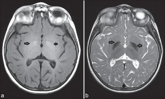
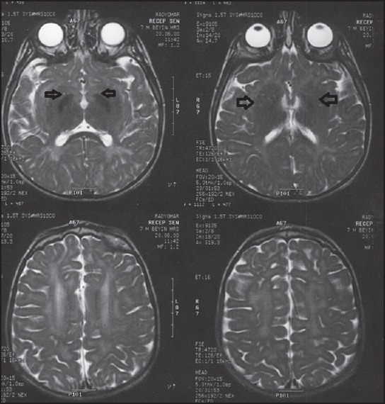
Fucosidosis is a rare lysosomal storage disease with clinical presentation of developmental retardation, coarse facial features, hepatosplenomegaly, dysostosis multiplex, and angiokeratomas. Here, a 7-year-old female patient with progressive dystonic movement disorder and loss of acquired motor skills is presented. Coarse facial feature and abnormal globuspallidus signaling in brain magnetic resonance imaging (MRI) led the patient to be investigated in terms of fucosidosis despite absence of hepatosplenomegaly, dysostosis multiplex, and angiokeratomas. Markedly decreased enzyme activity of alpha-fucosidosis led to the correct diagnosis.
Conclusion: Various neurological findings have recently been reported in fucosidosis. However, neuroimaging findings have not been studied in detail except a few studies. It is critically important to discuss the wide neuroradiological spectrum of the disease and to highlight fucosidosis in differential diagnosis of bilateral pallidalhypointensity on T2-weighted images in brain MRI. In addition, description of atypical clinical findings of fucosidosis should avoid clinicians from diagnostic delay.
Keywords: Dystonia; fucosidosis; globus pallidus hypointensity; magnetic resonance imaging.
Conflict of interest statement
Figures
Similar articles
-
Fucosidosis: A Review of a Rare Disease.Int J Mol Sci. 2025 Jan 3;26(1):353. doi: 10.3390/ijms26010353. Int J Mol Sci. 2025. PMID: 39796208 Free PMC article. Review.
-
Late diagnosis of fucosidosis in a child with progressive fixed dystonia, bilateral pallidal lesions and red spots on the skin.Eur J Paediatr Neurol. 2014 Jul;18(4):516-9. doi: 10.1016/j.ejpn.2014.02.005. Epub 2014 Feb 25. Eur J Paediatr Neurol. 2014. PMID: 24636010
-
Evolution of the neuroimaging changes in fucosidosis type II.J Inherit Metab Dis. 1996;19(6):775-81. doi: 10.1007/BF01799172. J Inherit Metab Dis. 1996. PMID: 8982951
-
MRI and MRS findings in fucosidosis; a rare lysosomal storage disease.Brain Dev. 2016 Apr;38(4):435-8. doi: 10.1016/j.braindev.2015.09.013. Epub 2015 Oct 26. Brain Dev. 2016. PMID: 26515723
-
A Novel Homozygous Mutation in the FUCA1 Gene Highlighting Fucosidosis as a Cause of Dystonia: Case Report and Literature Review.Neuropediatrics. 2019 Aug;50(4):248-252. doi: 10.1055/s-0039-1684052. Epub 2019 May 7. Neuropediatrics. 2019. PMID: 31064022 Review.
Cited by
-
Fucosidosis-Clinical Manifestation, Long-Term Outcomes, and Genetic Profile-Review and Case Series.Genes (Basel). 2020 Nov 22;11(11):1383. doi: 10.3390/genes11111383. Genes (Basel). 2020. PMID: 33266441 Free PMC article. Review.
-
Facial features of lysosomal storage disorders.Expert Rev Endocrinol Metab. 2022 Nov;17(6):467-474. doi: 10.1080/17446651.2022.2144229. Epub 2022 Nov 16. Expert Rev Endocrinol Metab. 2022. PMID: 36384353 Free PMC article.
-
Eye of the Tiger: Looking Beyond Neurodegeneration with Brain Iron Accumulation Disorders.J Pediatr Genet. 2021 Feb 17;12(2):163-166. doi: 10.1055/s-0041-1723959. eCollection 2023 Jun. J Pediatr Genet. 2021. PMID: 37090832 Free PMC article.
-
Fucosidosis: A Review of a Rare Disease.Int J Mol Sci. 2025 Jan 3;26(1):353. doi: 10.3390/ijms26010353. Int J Mol Sci. 2025. PMID: 39796208 Free PMC article. Review.
-
Diagnosis and Supportive Management of Fucosidosis: A Case Report.Cureus. 2019 Nov 12;11(11):e6139. doi: 10.7759/cureus.6139. Cureus. 2019. PMID: 31886074 Free PMC article.
References
-
- George HT. Scriver CR, Beaudet AL, Sly WS, Valle D, editors. Disorders of glycoprotein degradation: α-mannosidosis, β-mannosidosis, α-fucosidosis and sialidosis. Child B, Kinzler KW, Vogelstein B, assoc. 8th ed. The Metabolic and Molecular Basis of Inherited Metabolic Disease. 2001;3:3507–33.
-
- Durand P, Borrone C, Della Cella G, Philippart M. Fucosidosis. Lancet. 1968;1:1198. - PubMed
-
- Van Hoff F, Hers HG. Mucopolysaccharidosis by absence of α-fucosidase. Lancet. 1968;1:1198. - PubMed
-
- Willems PJ, Gatti R, Darby JK, Romeo G, Durand P, Dumon JE, et al. Fucosidosis revisited: A review of 77 patients. Am J Med Genet. 1991;38:111–31. - PubMed
Publication types
LinkOut - more resources
Full Text Sources
Other Literature Sources
