

CT Findings in People Who Were Environmentally Exposed to Asbestos in Korea
- PMID: 26713068
- PMCID: PMC4689837
- DOI: 10.3346/jkms.2015.30.12.1896
CT Findings in People Who Were Environmentally Exposed to Asbestos in Korea
Abstract
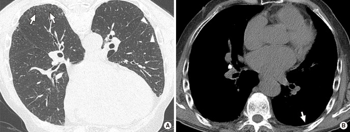
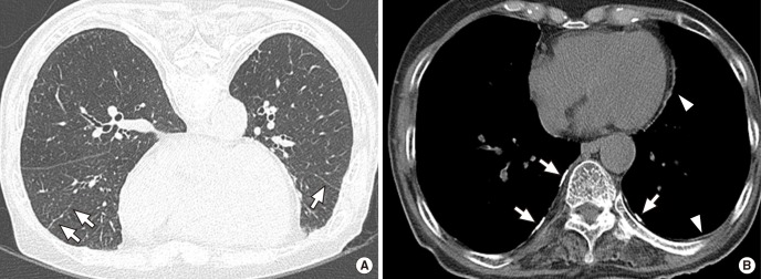
Asbestos related pleuropulmonary disease has been emerging health problem for recent years. It can cause variable clinical symptoms and radiological abnormalities. However, there has been no report for their characteristics in subjects who were environmentally exposed to asbestos. We reviewed the CT images of 35 people who were environmentally exposed to asbestos in Chungnam province, Korea. The study result showed high incidence of pleural plaque and pulmonary fibrosis on chest CT (94% and 77%, respectively). The common CT findings of lung parenchymal lesions were as follows: centrilobular opacities (94%), subpleural dot-like or branching opacities (80%), interlobular septal thickening (57%), intralobular interstitial thickening (46%), parenchymal bands (43%) and subpleural curvilinear line (29%). There were no significant differences in the prevalence of pulmonary fibrosis and pleural plaques according to sex, age and duration of exposure. In conclusion, pleural plaque and pulmonary fibrosis are common asbestos-related CT finding in the exposed people. Asbestos related lung parenchymal CT findings in the participants with environmental exposure show similar to those observed in the occupational exposure.
Keywords: Asbestos; Asbestosis; Environmental Exposure; Multidetector Computed Tomography; Plaque; Pulmonary Fibrosis.
Conflict of interest statement
Figures
Similar articles
-
[Pleural and parenchymal lung diseases from asbestos exposure. CT diagnosis].Radiol Med. 2000 Nov;100(5):326-31. Radiol Med. 2000. PMID: 11213409 Italian.
-
Asbestos-related pleuropulmonary diseases: evaluation with low-dose four-detector row spiral CT.Radiology. 2004 Oct;233(1):182-90. doi: 10.1148/radiol.2331031133. Epub 2004 Aug 27. Radiology. 2004. PMID: 15333769
-
Asbestosis, pleural plaques and diffuse pleural thickening: three distinct benign responses to asbestos exposure.Eur Respir J. 1998 May;11(5):1021-7. doi: 10.1183/09031936.98.11051021. Eur Respir J. 1998. PMID: 9648950
-
Asbestos: when the dust settles an imaging review of asbestos-related disease.Radiographics. 2002 Oct;22 Spec No:S167-84. doi: 10.1148/radiographics.22.suppl_1.g02oc10s167. Radiographics. 2002. PMID: 12376609 Review.
-
Computed tomography in the diagnosis of asbestos-related thoracic disease.J Thorac Imaging. 1989 Jan;4(1):61-7. doi: 10.1097/00005382-198901000-00012. J Thorac Imaging. 1989. PMID: 2643716 Review.
Cited by
-
The Asbestos Ban in Korea from a Grassroots Perspective: Why Did It Occur?Int J Environ Res Public Health. 2018 Jan 25;15(2):198. doi: 10.3390/ijerph15020198. Int J Environ Res Public Health. 2018. PMID: 29370079 Free PMC article.
-
Radiologic Diagnosis of Asbestosis in Korea.Korean J Radiol. 2016 Sep-Oct;17(5):674-83. doi: 10.3348/kjr.2016.17.5.674. Epub 2016 Aug 23. Korean J Radiol. 2016. PMID: 27587956 Free PMC article. Review.
-
Incidence and prevalence of interstitial lung diseases worldwide: a systematic literature review.BMJ Open Respir Res. 2023 Jun;10(1):e001291. doi: 10.1136/bmjresp-2022-001291. BMJ Open Respir Res. 2023. PMID: 37308252 Free PMC article.
References
-
- Silva CI, Müller NL. Asbestos-related disease. In: Müller NL, Silva CI, editors. Imaging of the chest. Philadelphia: Elsevier Saunders; 2008. pp. 1140–1166.
-
- Fraser RS, Colman N, Müller NL, Paré PD. Pulmonary disease caused by inhaled inorganic dust. In: Fraser RS, Colman N, Müller NL, Paré PD, editors. Synopsis of diseases of the chest. 3rd ed. Philadelphia: Elsevier Saunders; 2005. pp. 714–743.
-
- Goldberg P, Goldberg M, Marne MJ, Hirsch A, Tredaniel J. Incidence of pleural mesothelioma in New Caledonia: a 10-year survey (1978-1987) Arch Environ Health. 1991;46:306–309. - PubMed
Publication types
MeSH terms
Substances
LinkOut - more resources
Full Text Sources
Other Literature Sources
Medical
