

Intracranial Vasospasm without Intracranial Hemorrhage due to Acute Spontaneous Spinal Subdural Hematoma
- PMID: 26713084
- PMCID: PMC4688336
- DOI: 10.5607/en.2015.24.4.366
Intracranial Vasospasm without Intracranial Hemorrhage due to Acute Spontaneous Spinal Subdural Hematoma
Abstract
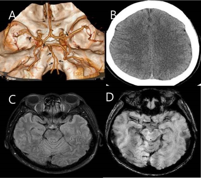
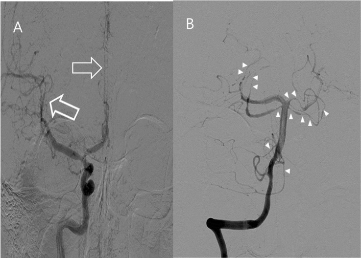
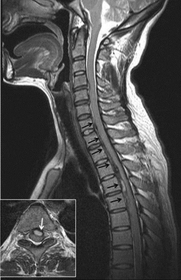
Spontaneous spinal subdural hematoma (SDH) is very rare. Furthermore, intracranial vasospasm (ICVS) associated with spinal hemorrhage has been very rarely reported. We present an ICVS case without intracranial hemorrhage following SDH. A 41-year-old woman was admitted to our hospital with a complaint of severe headache. Multiple intracranial vasospasms were noted on a brain CT angiogram and transfemoral cerebral angiography. However, intracranial hemorrhage was not revealed by brain MRI or CT. On day 3 after admission, weakness of both legs and urinary incontinence developed. Spine MRI showed C7~T6 spinal cord compression due to hyperacute stage of SDH. After hematoma evacuation, her symptoms gradually improved. We suggest that spinal cord evaluation should be considered in patients with headache who have ICVS, although intracranial hemorrhage would not be visible in brain images.
Keywords: Headache; Intracranial vasospasm; Spinal subdural hematoma; Subarachnoid hemorrhage.
Figures
Similar articles
-
Intracranial Vasospasm After Evacuation of Acute Spontaneous Subdural Hematoma.Cureus. 2021 May 27;13(5):e15284. doi: 10.7759/cureus.15284. Cureus. 2021. PMID: 34194885 Free PMC article.
-
Intracranial extension of spinal subarachnoid hematoma causing severe cerebral vasospasm.J Korean Neurosurg Soc. 2014 Dec;56(6):527-30. doi: 10.3340/jkns.2014.56.6.527. Epub 2014 Dec 31. J Korean Neurosurg Soc. 2014. PMID: 25628817 Free PMC article.
-
Spontaneous spinal and intracranial subdural hematoma.J Formos Med Assoc. 2009 Mar;108(3):258-61. doi: 10.1016/S0929-6646(09)60061-9. J Formos Med Assoc. 2009. PMID: 19293043
-
Concomitant Intracranial Chronic Subdural Hematoma and Spinal Subdural Hematoma: A Case Report and Literature Review.World Neurosurg. 2016 Jun;90:706.e1-706.e9. doi: 10.1016/j.wneu.2016.03.020. Epub 2016 Mar 18. World Neurosurg. 2016. PMID: 26996734 Review.
-
Aneurysmal Subarachnoid Hemorrhage with Spinal Subdural Hematoma: A Case Report and Systematic Review of the Literature.World Neurosurg. 2019 Aug;128:240-247. doi: 10.1016/j.wneu.2019.05.069. Epub 2019 May 17. World Neurosurg. 2019. PMID: 31103768
Cited by
-
Non-traumatic Spinal Subdural Hemorrhage Associated With Rivaroxaban Use.Cureus. 2024 Apr 28;16(4):e59208. doi: 10.7759/cureus.59208. eCollection 2024 Apr. Cureus. 2024. PMID: 38807840 Free PMC article.
-
Intracranial Vasospasm After Evacuation of Acute Spontaneous Subdural Hematoma.Cureus. 2021 May 27;13(5):e15284. doi: 10.7759/cureus.15284. Cureus. 2021. PMID: 34194885 Free PMC article.
-
Report of cerebral vasospasm as a complication of intracranial subarachnoid hemorrhage following traumatic lumbar puncture.Surg Neurol Int. 2022 Apr 8;13:128. doi: 10.25259/SNI_181_2022. eCollection 2022. Surg Neurol Int. 2022. PMID: 35509586 Free PMC article.
-
Acute spontaneous spinal subdural hematoma presenting with Takotsubo cardiomyopathy: a rare case report and literature review.EFORT Open Rev. 2022 May 5;7(5):312-317. doi: 10.1530/EOR-22-0003. EFORT Open Rev. 2022. PMID: 35510739 Free PMC article. Review.
References
-
- Findlay JM, Nisar J, Darsaut T. Cerebral vasospasm: a review. Can J Neurol Sci. (in press) - PubMed
-
- Domenicucci M, Ramieri A, Ciappetta P, Delfini R. Nontraumatic acute spinal subdural hematoma: report of five cases and review of the literature. J Neurosurg. 1999;91:65–73. - PubMed
-
- Kyriakides AE, Lalam RK, El Masry WS. Acute spontaneous spinal subdural hematoma presenting as paraplegia: a rare case. Spine (Phila Pa 1976) 2007;32:E619–E622. - PubMed
-
- Yoon CD, Song CJ, Lee JE, Choi SW. Simultaneous intracranial and spinal subdural hematoma: two case reports. J Korean Soc Radiol. 2009;60:79–82.
Publication types
LinkOut - more resources
Full Text Sources
Other Literature Sources
Miscellaneous
