Osteoporotic Fracture: 2015 Position Statement of the Korean Society for Bone and Mineral Research
- PMID: 26713308
- PMCID: PMC4691591
- DOI: 10.11005/jbm.2015.22.4.175
Osteoporotic Fracture: 2015 Position Statement of the Korean Society for Bone and Mineral Research
Abstract
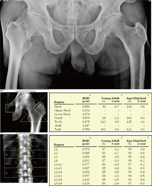
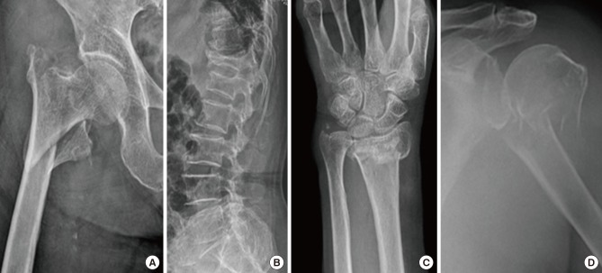
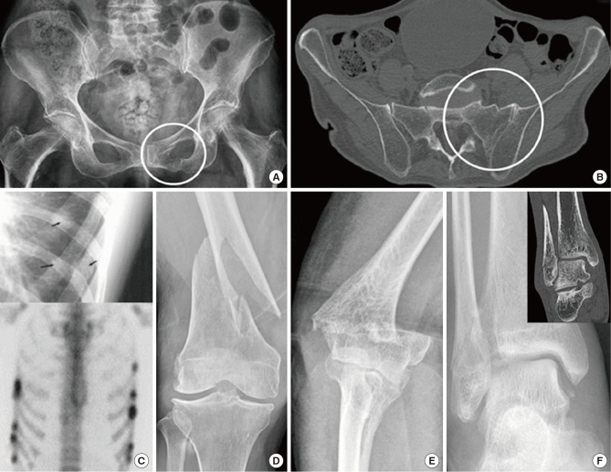
Osteoporotic fractures are one of the most common causes of disability and a major contributor to medical care costs worldwide. Prior osteoporotic fracture at any site is one of the strongest risk factors for a new fracture, which occurs very soon after the first fracture. Bone mineral density (BMD) scan, a conventional diagnostic tool for osteoporosis, has clear limitations in diagnosing osteoporotic fractures and identifying the risk of subsequent fractures. Therefore, early and accurate diagnosis of osteoporotic fractures using the clinical definition which is applicable practically and independent of BMD, is essential for preventing subsequent fractures and reducing the socioeconomic burden of these fractures. Fractures caused by low-level trauma equivalent to a fall from a standing height or less at major (hip, spine, distal radius, and proximal humerus) or minor (pelvis, sacrum, ribs, distal femur and humerus, and ankle) sites in adults over age 50, should be first regarded as osteoporotic. In addition, if osteoporotic fractures are strongly suspected on history and physical examination even though there are no positive findings on conventional X-rays, more advanced imaging techniques such as computed tomography, bone scan, and magnetic resonance imaging are necessary as soon as possible.
Keywords: Definition; Diagnosis; Osteoporosis; Osteoporotic fractures; Practice guidelines as topic.
Conflict of interest statement
No potential conflict of interest relevant to this article was reported.
Figures
References
-
- Schuit SC, van der Klift M, Weel AE, et al. Fracture incidence and association with bone mineral density in elderly men and women: the Rotterdam Study. Bone. 2004;34:195–202. - PubMed
-
- Johnell O, Kanis JA. An estimate of the worldwide prevalence and disability associated with osteoporotic fractures. Osteoporos Int. 2006;17:1726–1733. - PubMed
-
- Lee KM, Chung CY, Kwon SS, et al. Ankle fractures have features of an osteoporotic fracture. Osteoporos Int. 2013;24:2819–2825. - PubMed
Publication types
LinkOut - more resources
Full Text Sources
Other Literature Sources