. 2016;15(3):413-24.
doi: 10.1080/15384101.2015.1127472.

Aurora A phosphorylation of WD40-repeat protein 62 in mitotic spindle regulation

Affiliations

Aurora A phosphorylation of WD40-repeat protein 62 in mitotic spindle regulation

Nicholas R Lim et al. Cell Cycle. 2016.

Abstract

Mitotic spindle organization is regulated by centrosomal kinases that potentiate recruitment of spindle-associated proteins required for normal mitotic progress including the microcephaly protein WD40-repeat protein 62 (WDR62). WDR62 functions underlie normal brain development as autosomal recessive mutations and wdr62 loss cause microcephaly. Here we investigate the signaling interactions between WDR62 and the mitotic kinase Aurora A (AURKA) that has been recently shown to cooperate to control brain size in mice. The spindle recruitment of WDR62 is closely correlated with increased levels of AURKA following mitotic entry. We showed that depletion of TPX2 attenuated WDR62 localization at spindle poles indicating that TPX2 co-activation of AURKA is required to recruit WDR62 to the spindle. We demonstrated that AURKA activity contributed to the mitotic phosphorylation of WDR62 residues Ser49 and Thr50 and phosphorylation of WDR62 N-terminal residues was required for spindle organization and metaphase chromosome alignment. Our analysis of several MCPH-associated WDR62 mutants (V65M, R438H and V1314RfsX18) that are mislocalized in mitosis revealed that their interactions and phosphorylation by AURKA was substantially reduced consistent with the notion that AURKA is a key determinant of WDR62 spindle recruitment. Thus, our study highlights the role of AURKA signaling in the spatiotemporal control of WDR62 at spindle poles where it maintains spindle organization.

Keywords: aurora A; c-jun N-terminal kinase; mitosis; phosphorylation; primary microcephaly.


Figures

Figure 1.
Figure 1.
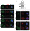
WDR62 deletion by CRISPR/Cas9-sgRNA does not alter mitotic AURKA expression or phosphorylation. (A) Genomic sequence verification of a single-base insertion leading to a frame-shift deletion of the wdr62 gene. (B) AD293 (5×10⁶) cells deleted of WDR62 (WDR62 sgRNA), unedited controls (control) and parental cells (AD293) were immunoblotted to determine WDR62 expression levels. Mean values are of densitometric measurements of mitotic pAURKA, AURKA and TPX2 bands normalized against tubulin loading. (n=3, error bars indicate SEM, ‘ns’ – not significant, P > 0.05, students t-test) (C) WDR62-deleted AD293 cells (KO) or unedited controls (con) were immunostained to evaluate WDR62 expression and localization on the mitotic spindle. D) WDR62-deleted AD293 cells (KO) or unedited controls (con) synchronised in mitosis (Noc, 350 nM, 16 h) or asynchronous (Async) were immunoblotted to determine AURKA expression/phosphorylation, TPX2 levels and with mitotic markers (Cyclin B1, pHH3). Quantitations are from 3 independent repeated densitometric measure of AURKA phosphorylation and expression and normalized against αtubulin expression as a loading control. (E) WDR62-deleted AD293 cells (KO) or unedited controls (control) were immunostained to evaluate the localization of total and active AURKA on the mitotic spindle. All scale bars represent 10 µm.
Figure 2.
Figure 2.
WDR62 spindle pole localization in mitosis is dependent on TPX2 expression. (A) Hela cells were fixed and the localization of endogenous WDR62 and AURKA determined at different stages of mitosis by immunofluorescence. Scale bars represent 10 µm. (B) HeLa cells transfected with TPX2 siRNA (siTPX2) or non-targeting control siRNA (siCon) were synchronized in mitosis (Noc, 350 nM, 16 h) before immunoblot analysis to confirm depletion of TPX2 expression and reduction of phosphorylated AURKA. AURKA or WDR62 expression levels were also evaluated. (C) HeLa cells transfected with TPX2 siRNA or non-targeting control siRNA (siCon) and the localization and expression of TPX2, AURKA and WDR62 determined by co-immunofluorecence staining. Scale bars represent 10 µm.
Figure 3.
Figure 3.
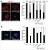
AURKA phosphorylation of WDR62 is required for metaphase spindle regulation and chromosomal alignment. (A) WDR62-deleted AD293 cells (KO) were transfected with wild-type WDR62 (WT), Ser32/Ser33A (32/33A), Ser49/Thr50/Ser52A (49/50/52A), or Ser32/Ser33/Ser49/Thr50/Ser52A (5A) mutant, fixed and stained with γ-tubulin for metaphase spindle phenotype analysis (graphed). Representative images of spindle defects (multipolar, abnormal and monopolar) are shown on the left panels. (B) WDR62-deleted cells (KO) were transfected with wild-type WDR62 (WT), Ser32/Ser33A (32/33A), Ser49/Thr50/Ser52A (49/50/52A), or 5A mutant, fixed and stained with DAPI to analyze for chromosomal alignment defects (graphed). Representative images of normal and misaligned chromosomes in metaphase are shown on the left panels. “n” indicates number of cells scored. Scale bars represent 10 µm.
Figure 4.
Figure 4.
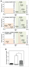
Mitotic phosphorylation of WDR62 on Ser49 and Thr50 is dependent on AURKA activity. (A) WDR62 knockout cells (1×10⁸ cells) were transfected with wild-type WDR62 and synchronised with hydroxyurea (top panel), nocodazole (middle panel), or synchronised with nocodazole and treated with MLN8237 (bottom panel). Cells were then lysed, trypsin-digested, spiked with equal amounts of synthetic peptide standard 46TRL(p)s(p)tASEETVQNR(13C15N)58, phospho-enriched, before quantitation using LC-MS/MS (n=3). Representative spectra of the endogenous and spiked synthetic peptides were obtained from the chromatographic peak apex. (B) Quantification of relative abundance of wild-type 46TRL(p)s(p)tASEETVQN58 peptide in S phase, M phase, and M phase with MLN8237-treated AD293 cells from (A). (n=3, error bars indicate SEM *P < 0.05, students t-test).
Figure 5.
Figure 5.
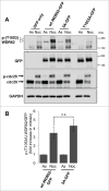
AURKA signaling is not required for WDR62 T1053 phosphorylation in mitosis. (A) AD293 (WDR62 KO) cells transiently expressing GFP-tagged wild-type WDR62, WDR62-5A, WDR62 T1053A mutants or a vector expressing GFP only, Mitotically synchronized (Noc, 350 nM, 16 h) or asynchronous (As) cells were lysed and immunoblotted with a site specific phospho-(T1053) WDR62 antibody. GFP-fusion protein expression, mitotic status (cdc25 phosphorylation-dependent band shift) and protein loading (GAPDH) was also determined by immunoblotting. (B) Densitometric measurements of phospho-(T1053) WDR62 bands from were normalized for GFP expression and expressed as fold increases over asynchronous cells (As). Values represent mean + SEM (n=3, ‘ns’ – not significant, p > 0.05, students t-test).
Figure 6.
Figure 6.
AURKA signaling to WDR62 is disrupted by MCPH mutations. (A) WDR62 deleted AD293 (WDR62 KO) cells were transfected with empty myc vector, myc-tagged wild-type WDR62 (wt-WDR62) or myc-tagged MCPH-associated WDR62 mutants (V65M, R438H and V1314RfsX18). Mitotically synchronized (Noc, 350 nM, 16 h) cell lysates were then immunoprecipitated with anti-myc and blotted for AURKA. (B) AD293 cells were transfected with WDR62 V65M, R438H or V1314RfsX18 mutants or wild-type WDR62 (wt-WDR62) , synchronised in S (hydroxyurea, 5 mM, 16 h) or M phase (Noc, 350 nM, 16 h), followed by quantitative phosphopeptide analysis by LC-MS/MS in presence of the spiked synthetic peptides. Representative extracted ion chromatogram of the 46TRL(p)s(p)tASEETVQN58 peptide in these samples are shown. Peaks corresponding to the peptide is highlighted in blue (n=3). (C) Asynchronous (As) or mitotically synchronized (Noc) WDR62 KO cells transiently expressing GFP-tagged WDR62 (wt-WDR62) or MCPH-associated WDR62 mutants (V65M, R438H and V1314RfsX18) were immunoblotted to determine WDR62 T1053 phosphorylation or GFP-fusion protein expression.

Comment in

  • Driving WDR62 to the pole.
    Xu D, Xu Z. Xu D, et al. Cell Cycle. 2016 May 2;15(9):1180-1. doi: 10.1080/15384101.2016.1154374. Epub 2016 Apr 25. Cell Cycle. 2016. PMID: 27110649 Free PMC article. No abstract available.

References

    1. Lampson MA, Renduchitala K, Khodjakov A, Kapoor TM. Correcting improper chromosome-spindle attachments during cell division. Nat Cell Biol 2004; 6:232-37; PMID:14767480; http://dx.doi.org/10.1038/ncb1102 - DOI - PubMed
    1. Compton DA. Spindle assembly in animal cells. Annu Rev Biochem 2000; 69:95-114; PMID:10966454; http://dx.doi.org/10.1146/annurev.biochem.69.1.95 - DOI - PubMed
    1. Zimmerman WC, Sillibourne J, Rosa J, Doxsey SJ. Mitosis-specific anchoring of gamma tubulin complexes by pericentrin controls spindle organization and mitotic entry. Mol Biol Cell 2004; 15:3642-57; PMID:15146056; http://dx.doi.org/10.1091/mbc.E03-11-0796 - DOI - PMC - PubMed
    1. Oshimori N, Li X, Ohsugi M, Yamamoto T. Cep72 regulates the localization of key centrosomal proteins and proper bipolar spindle formation. EMBO J 2009; 28:2066-76; PMID:19536135; http://dx.doi.org/10.1038/emboj.2009.161 - DOI - PMC - PubMed
    1. Bornens M. The centrosome in cells and organisms. Science 2012; 335:422-6; PMID:22282802; http://dx.doi.org/10.1126/science.1209037 - DOI - PubMed

Publication types

MeSH terms