Review
. 2016 Jan;19(1):10-9.
doi: 10.1038/nn.4190.

An epigenetic gateway to brain tumor cell identity

Affiliations
Review

An epigenetic gateway to brain tumor cell identity

Stephen C Mack et al. Nat Neurosci. 2016 Jan.

Abstract

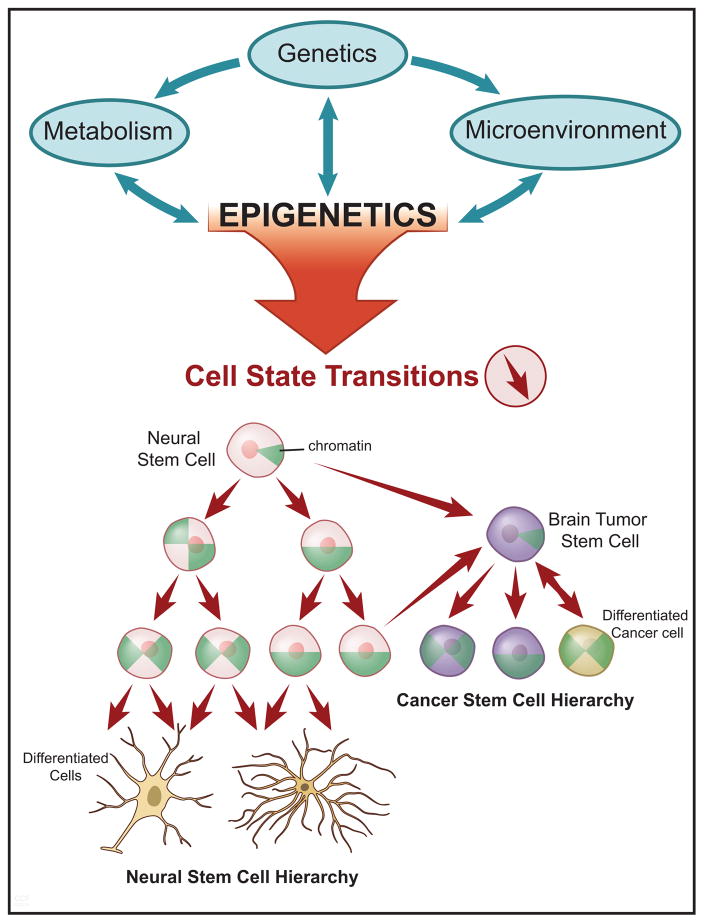
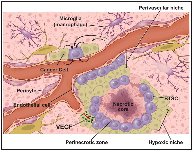
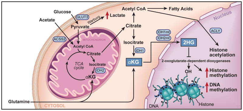
Precise targeting of genetic lesions alone has been insufficient to extend brain tumor patient survival. Brain cancer cells are diverse in their genetic, metabolic and microenvironmental compositions, accounting for their phenotypic heterogeneity and disparate responses to therapy. These factors converge at the level of the epigenome, representing a unified node that can be disrupted by pharmacologic inhibition. Aberrant epigenomes define many childhood and adult brain cancers, as demonstrated by widespread changes to DNA methylation patterns, redistribution of histone marks and disruption of chromatin structure. In this Review, we describe the convergence of genetic, metabolic and microenvironmental factors on mechanisms of epigenetic deregulation in brain cancer. We discuss how aberrant epigenetic pathways identified in brain tumors affect cell identity, cell state and neoplastic transformation, as well as addressing the potential to exploit these alterations as new therapeutic strategies for the treatment of brain cancer.


Conflict of interest statement

Conflict of Interest

The authors declare no conflict of interest.

Figures

Figure 1
Figure 1. The Epigenetic Gateway to Cell Identity and Neoplastic Transformation
A schematic depicting the genetic, metabolic, and microenvironmental interactions (green arrows) with epigenetic programs in cancer (top panel). In the lower panel, a diagram illustrating the cell state transitions (red arrows) influenced by altered epigenetic landscapes and their relevance to both normal neural stem cell, and cancer stem cell hierarchies (lower panel). Within the cells are green pie-shaped triangles, which represent the restructuring of chromatin architecture and progression towards closed chromatin in the most differentiated cell state.
Figure 2
Figure 2. Brain Tumors Converge on Chromatin Architecture
A diagram depicting euchromatin and histone modifications that mediate ‘active’ transcription in cancer cells (top panel). Shown are various histone modifications and enzymes, which catalyze the addition of post-translational modifications such as histone methylation and acetylation, or bind to these modifications, such as the BRD4, which binds acetylated lysine residues on histones. The green ovals represent transcription factor binding sites and locations of enhancers, or clusters of enhancers, termed super-enhancers. Also shown are drug compounds, which inhibit the removal (Vorinostat and Pabinostat - Histone deacetylase (HDAC) inhibitor) or detection of acetylation (JQ1). Shown in the middle are the chromatin remodelers, which facilitate the landscape of higher order chromatin structure towards euchromatin or heterochromatin. In the lower panel, heterochromatin is depicted and the associated modifications that mediate tumor suppressor gene silencing. These include the DNA methyltransferase family of enzymes, which catalyze the addition of methyl groups to cytosine - guanine di-nuclueotides, and TET enzymes, which facilitate DNA de-methylation through 5-methyl cytosine hydroxylation. Also shown is EZH2, which methylates at histone H3 at the 27th position, and the associated histone H3K27 de-methylases KDM6A and JMJD3. Shown are chemical inhibitors that reverse the methylation marks deposited or removed by these methyltransferase and demethylase enzymes related to heterochromatin (Decitabine, GSK343, GSKJ4). ATRX and DAXX are depicted which function to incorporate the histone H3.3 variant, and which are frequently mutated in pediatric high-grade glioma.
Figure 3
Figure 3. Cellular Microenvironment Influences Epigenetic State of Brain Tumor Cells
An illustration of the brain tumor microenvironment highlighting the perivascular and hypoxic niches, which dictate interacting cell types and nutrient availabilities. Both cancer cells (light green) and brain tumor cells (purple-round) exist in dynamic microenvironments containing exogenous signals from surrounding microglia (purple), pericytes (dark pink), endothelial cells (light pink), and other neoplastic cells. These interactions occur in the presence of variable growth factor gradients (ie. VEGF), oxygen availability, and nutrient levels (i.e. glucose, acetate, glutamine etc.).
Figure 4
Figure 4. Cellular Metabolism Influences Brain Cancer Epigenetic State
A schematic of metabolic pathways present within a bran tumor cell with emphasis on transport proteins (GLUT3), and enzymatic effectors (IDH1/2 mutations, ACSS2, and ACLY (ATP citrate lyase)), which alter tumor metabolism and ultimately epigenetic programs. The result of the IDH1 mutation is emphasized, which results in the accumulation of 2-hydroxyglutarate, a metabolite that inhibits the function of iron, oxygen, and α-ketoglutarate dependent demethylase enzymes, thus leading to aberrant accumulation of both DNA and histone methylation.

References

    1. Louis DN, et al. The 2007 WHO classification of tumours of the central nervous system. Acta neuropathologica. 2007;114:97–109. - PMC - PubMed
    1. Lawrence MS, et al. Discovery and saturation analysis of cancer genes across 21 tumour types. Nature. 2014;505:495–501. - PMC - PubMed
    1. Bettegowda C, et al. Exomic sequencing of four rare central nervous system tumor types. Oncotarget. 2013;4:572–583. - PMC - PubMed
    1. Jiao Y, et al. Frequent ATRX, CIC, FUBP1 and IDH1 mutations refine the classification of malignant gliomas. Oncotarget. 2012;3:709–722. - PMC - PubMed
    1. Parsons DW, et al. An integrated genomic analysis of human glioblastoma multiforme. Science. 2008;321:1807–1812. - PMC - PubMed

Publication types