. 2015 Dec 29;10(12):e0145512.
doi: 10.1371/journal.pone.0145512. eCollection 2015.

Effectiveness of Losartan-Loaded Hyaluronic Acid (HA) Micelles for the Reduction of Advanced Hepatic Fibrosis in C3H/HeN Mice Model

Affiliations

Effectiveness of Losartan-Loaded Hyaluronic Acid (HA) Micelles for the Reduction of Advanced Hepatic Fibrosis in C3H/HeN Mice Model

Reju George Thomas et al. PLoS One. .

Abstract

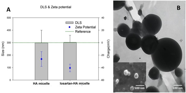
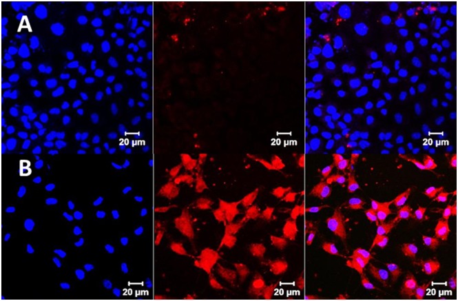
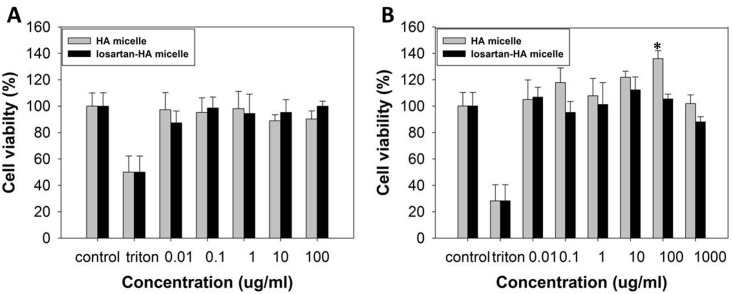
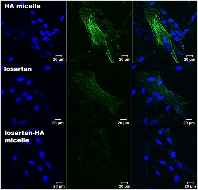
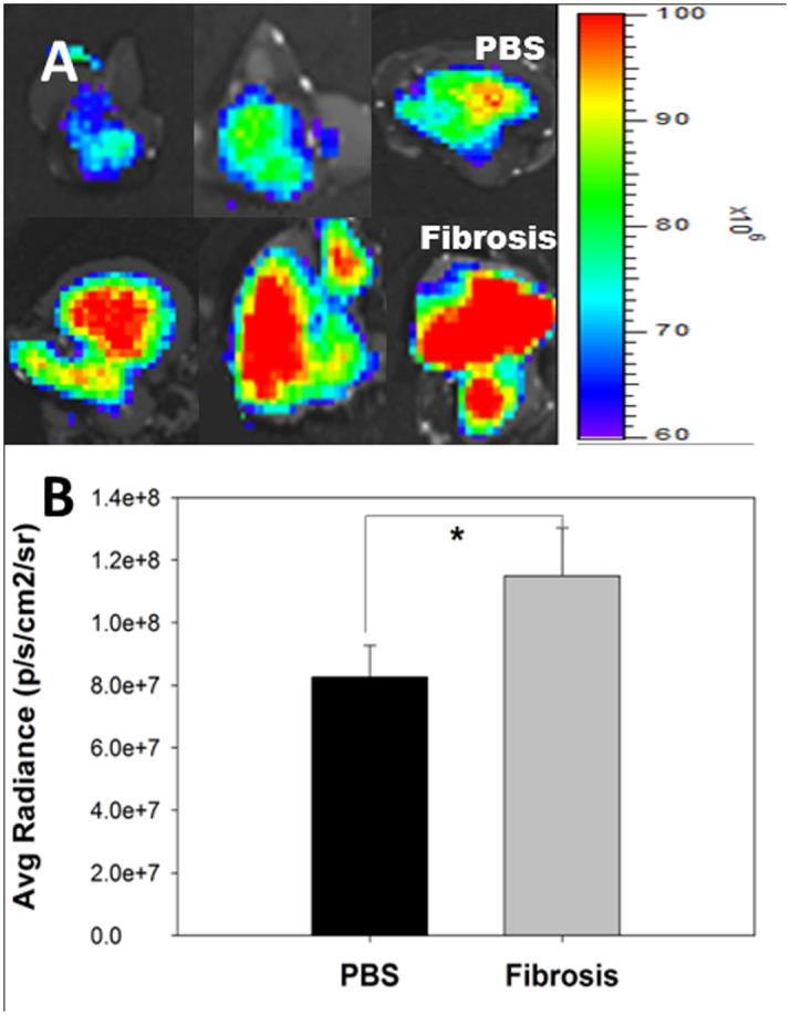
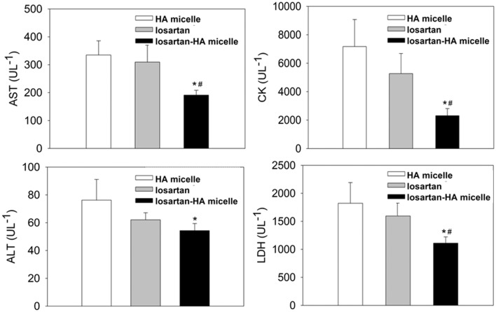
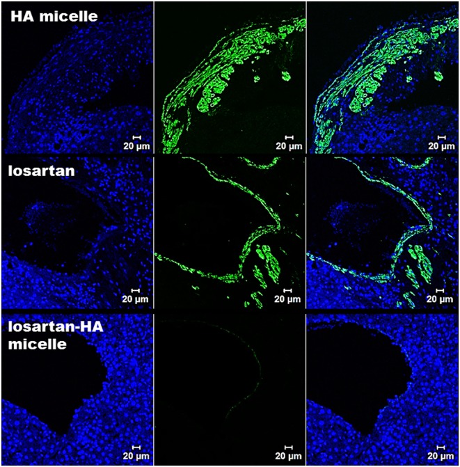
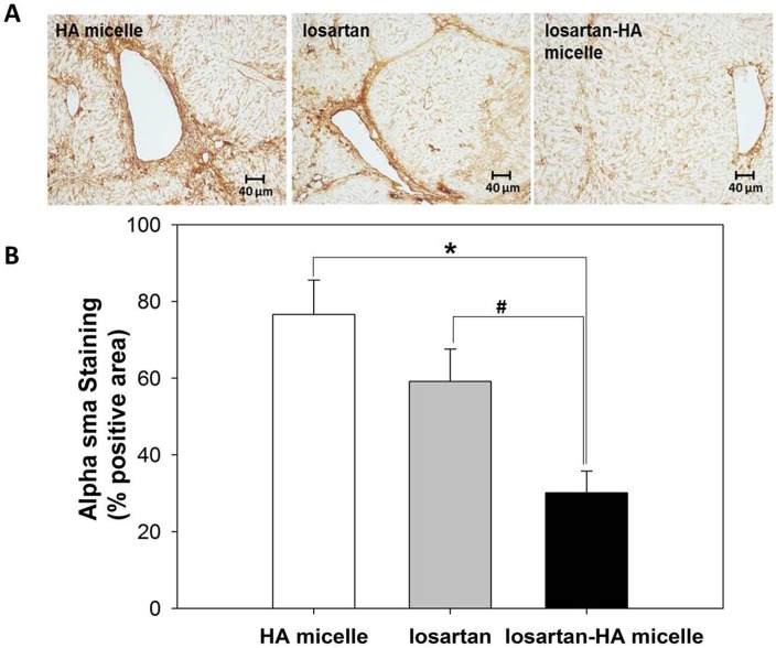
Advanced hepatic fibrosis therapy using drug-delivering nanoparticles is a relatively unexplored area. Angiotensin type 1 (AT1) receptor blockers such as losartan can be delivered to hepatic stellate cells (HSC), blocking their activation and thereby reducing fibrosis progression in the liver. In our study, we analyzed the possibility of utilizing drug-loaded vehicles such as hyaluronic acid (HA) micelles carrying losartan to attenuate HSC activation. Losartan, which exhibits inherent lipophilicity, was loaded into the hydrophobic core of HA micelles with a 19.5% drug loading efficiency. An advanced liver fibrosis model was developed using C3H/HeN mice subjected to 20 weeks of prolonged TAA/ethanol weight-adapted treatment. The cytocompatibility and cell uptake profile of losartan-HA micelles were studied in murine fibroblast cells (NIH3T3), human hepatic stellate cells (hHSC) and FL83B cells (hepatocyte cell line). The ability of these nanoparticles to attenuate HSC activation was studied in activated HSC cells based on alpha smooth muscle actin (α-sma) expression. Mice treated with oral losartan or losartan-HA micelles were analyzed for serum enzyme levels (ALT/AST, CK and LDH) and collagen deposition (hydroxyproline levels) in the liver. The accumulation of HA micelles was observed in fibrotic livers, which suggests increased delivery of losartan compared to normal livers and specific uptake by HSC. Active reduction of α-sma was observed in hHSC and the liver sections of losartan-HA micelle-treated mice. The serum enzyme levels and collagen deposition of losartan-HA micelle-treated mice was reduced significantly compared to the oral losartan group. Losartan-HA micelles demonstrated significant attenuation of hepatic fibrosis via an HSC-targeting mechanism in our in vitro and in vivo studies. These nanoparticles can be considered as an alternative therapy for liver fibrosis.


Conflict of interest statement

Competing Interests: This research was supported by Basic Research Program through the National Research Foundation of Korea (NRF) funded by the Ministry of Science and future Planning (2015R1A2A2A01007798). The funder (DKC Corporation (BioActs)) provided support in the form of salaries for authors and/or research materials [MMJ], but did not have any additional role in the study design, data collection and analysis, decision to publish, or preparation of the manuscript. The specific role of this author is articulated in the ‘author contributions’ section. There are no competing interests and this does not alter the authors' adherence to PLOS ONE policies on sharing data and materials.

Figures

Fig 1
Fig 1. Physicochemical characteristics of HA micelle and losartan-HA micelle.
A. Size measurement by DLS and surface charge measurement by zeta potential of HA micelle and losartan-HA micelle in PBS at 7.4 pH. HA micelle and losartan-HA micelle was taken at 100 μg/ml concentration and bath sonicated (Power Sonic410,Hwashin,Korea) for 5 minutes at room temperature and measured; B. TEM image of a SPION loaded HA micelle and SEM image of losartan-HA micelle (inset). The size of losartan-HA micelle was estimated to be around 300nm based on SEM and DLS result whereas the morphology of particle was confirmed from TEM and SEM image.
Fig 2
Fig 2. Cell uptake study of HA micelle labeled with Flamma552 dye by confocal microscopy.
A. Representative picture of FL83B cells incubated in 70 μg/ml of HA micelle labeled with Flamma552. Accumulation of HA micelle labeled with Flamma552 in FL83B cells was found to be minimal; B. Representative picture of hHSC cells incubated with 70 μg/ml of HA micelle labeled with Flamma552. HA micelle labeled with Flamma552 is clearly seen in HSC with more fluorescent intensity than FL83B. The incubation period was 2 hours for all experiments. Blue color represent uptake of DAPI stain in nucleus.
Fig 3
Fig 3. In vitro cytocompatibiliy of HA micelle and losartan-HA micelle (in PBS, pH 7.4).
A. hHSC; B. FL83B cell line. MTS assay of HA micelle and losartan-HA micelle has minimum toxicity effect even at high concentration (100 and 1,000 μg/ml). MTS assay data show the mean cell viability of quadruplicate samples ± SD. Losartan-HA micelle at 100 μg/ml showed significantly higher cell viability (*P <0.01) relative to the control in FL83B cell line. All other treatment does not show significant change in cell viability compared to control in both hHSC and FL83B cell lines (P>0.05).
Fig 4
Fig 4. Confocal microscopy imaging of α-sma expression in hHSC cell.
In HA micelle group, hHSC cells were incubated for 24 hours with HA micelle prior to 2 hour incubation with 1,000 nM of angiotensin 2. When angiotensin 2 is taken up by cells via angiotensin 1 receptor mechanism, expression of α-sma indicate the possible activation of HSC. In losartan group hHSC cells were incubated for 24 hours with 1,000nM losartan prior to 2 hour incubation with 1,000 nM of angiotensin 2. The angiotensin 1 receptors have been blocked by losartan which minimized expression of α-sma. In losartan-HA micelle group, hHSC cells were incubated for 24 hours with 1,000nM losartan-HA micelle prior to 2 hour incubation with 1,000 nM of angiotensin 2. Losartan-HA micelle helped in suppressing expression of α-sma more effectively. Blue color indicate DAPI stain.
Fig 5
Fig 5. Ex vivo profile of HA micelle labelled with with Flamma 774 in the liver.
(A) PBS treated normal mice shows accumulation of HA micelle labelled with with Flamma 774 (Top). TAA/Ethanol treated mice with liver fibrosis shows marked increased accumulation of HA micelle labelled with with Flamma 774 by CD44 receptor mediated uptake in HSC (Bottom). (B) Fluorescence intensity was quantified by the region-of-interest (ROI) method. The data are presented as the mean ± SEM. *P <0.05 relative to the liver ROI.
Fig 6
Fig 6. Blood biochemical estimation of AST, ALT, CK and LDH of HA micelle, losartan and losartan-HA micelle treated mice group after 20 weeks fibrosis induction by TAA/Ethanol treatment.
Losartan-HA micelle group have statistically significant lower AST/ALT, CK and LDH values compared to HA micelle and losartan group which indicate better treatment effect due to successful losartan delivery to hepatic stellate cells in liver. The data are presented as the mean ± SEM. *P <0.05 and #P <0.05 relative to HA micelle and losartan group, respectively.
Fig 7
Fig 7. Confocal microscopy imaging of α-sma expression in liver section.
In HA-micelle group, mice injected with HA micelle shows high expression of α-sma as the fibrotic state of liver have increased proliferation of activated HSC which express α-sma (represented by green fluorescence). In losartan group mice injected with losartan reveals similar expression profile of α-sma compared to HA micelle treated mice group as the oral losartan have failed to reach the target region (activated HSC). In losartan-HA micelle group, mice injected with losartan-HA micelle have almost no expression of α-sma which indicate successful delivery of losartan encapsulated losartan-HA micelle in the target region. Blue color indicate DAPI stain.
Fig 8
Fig 8. Immunohistochemical analysis of α-sma expression in liver section.
(A) Representative images of smooth muscle actin immunohistochemical staining in liver. (B) The expression of α-sma in losartan-HA micelle group is significantly lower than those of the HA micelle and losartan groups. The proportion of areas immunopositive for smooth muscle actin was measured using IHC profiler plugin in ImageJ (NIH image program, version 1.49) software as mean percent positive areas. The data are presented as the mean ± SD *P <0.001 and #P <0.001 relative to HA micelle and losartan group respectively.

References

    1. Hernandez-Gea V, Friedman SL. Pathogenesis of liver fibrosis. Annu Rev Pathol 2011;6: 425–456 10.1146/annurev-pathol-011110-130246 - DOI - PubMed
    1. Bosetti C, Levi F, Lucchini F, Zatonski WA, Negri E, La Vecchia C. Worldwide mortality from cirrhosis: an update to 2002. J Hepatol 2007;46:827–839. - PubMed
    1. Rehm J, Samokhvalov AV, Shield KD. Global burden of alcoholic liver diseases. J Hepatol 2013;59:160–168. 10.1016/j.jhep.2013.03.007 - DOI - PubMed
    1. Bataller R, Sancho-bru P, Ginès P, Lora JM, Al-garawi A, Solé M, et al. Activated human hepatic stellate cells express the renin-angiotensin system and synthesize angiotensin II. Gastroenterology. 2003;125:117–25. - PubMed
    1. Colmenero J, Bataller R, Sancho-Bru P, Domínguez M, Moreno M, Forns X, et al. Effects of losartan on hepatic expression of nonphagocytic NADPH oxidase and fibrogenic genes in patients with chronic hepatitis C. Am. J. Physiol., 2009: G726–G734. - PMC - PubMed

Publication types

MeSH terms