. 2015 Dec 29:4:e11897.
doi: 10.7554/eLife.11897.

Protein translocation channel of mitochondrial inner membrane and matrix-exposed import motor communicate via two-domain coupling protein

Affiliations

Protein translocation channel of mitochondrial inner membrane and matrix-exposed import motor communicate via two-domain coupling protein

Rupa Banerjee et al. Elife. .

Abstract

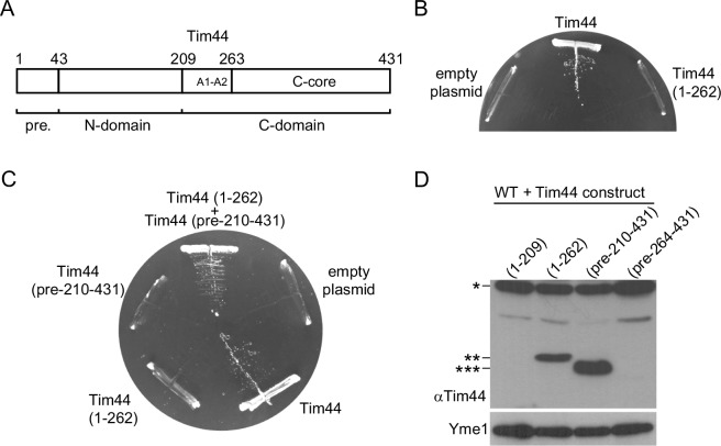
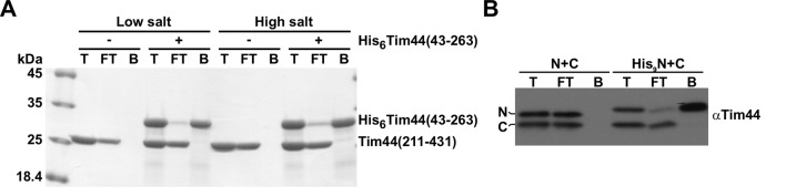
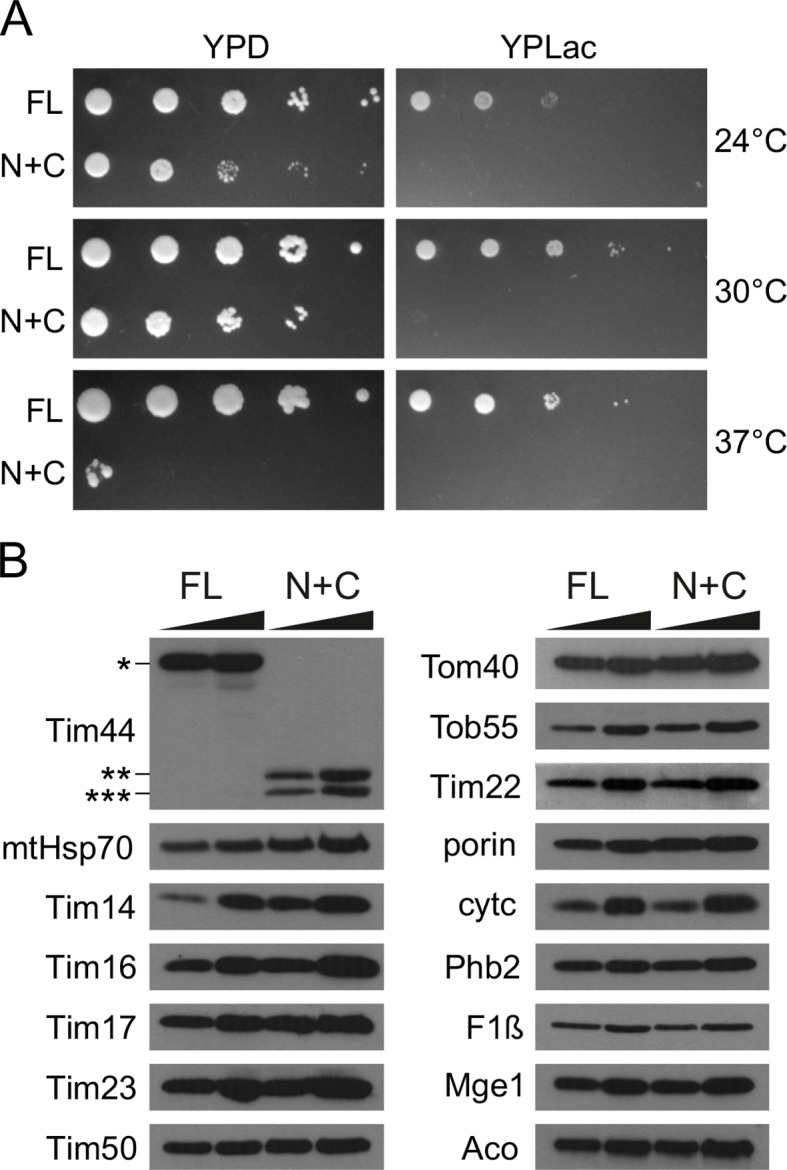
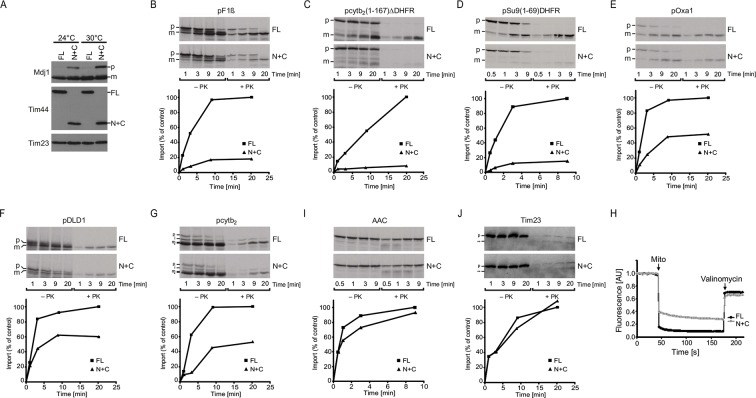
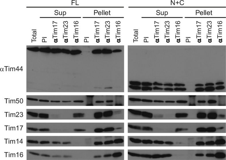
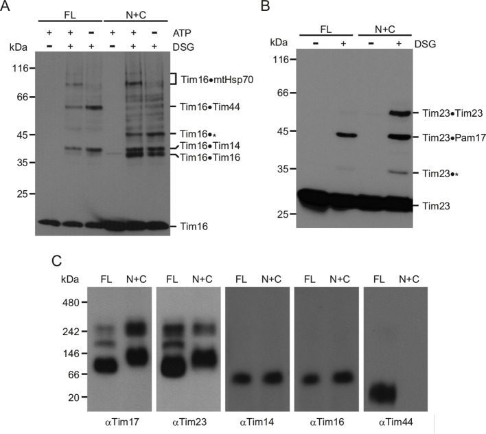
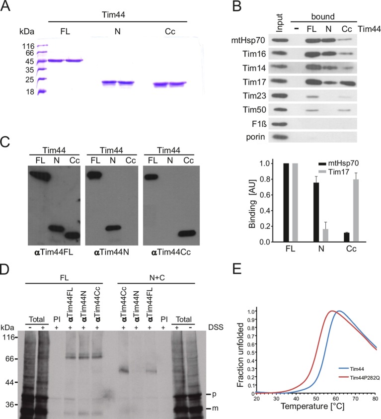
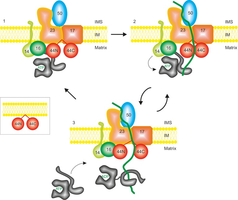
The majority of mitochondrial proteins are targeted to mitochondria by N-terminal presequences and use the TIM23 complex for their translocation across the mitochondrial inner membrane. During import, translocation through the channel in the inner membrane is coupled to the ATP-dependent action of an Hsp70-based import motor at the matrix face. How these two processes are coordinated remained unclear. We show here that the two domain structure of Tim44 plays a central role in this process. The N-terminal domain of Tim44 interacts with the components of the import motor, whereas its C-terminal domain interacts with the translocation channel and is in contact with translocating proteins. Our data suggest that the translocation channel and the import motor of the TIM23 complex communicate through rearrangements of the two domains of Tim44 that are stimulated by translocating proteins.

Keywords: E. coli; Hsp70; S. cerevisiae; TIM23; biochemistry; cell biology; mitochondria; protein translocation/sorting.


Conflict of interest statement

The authors declare that no competing interests exist.

Figures

Figure 1.
Figure 1.. The function of Tim44 can be rescued by its two domains expressed in trans but not by either of the domains alone.
(A) Schematic representation of Tim44 domain structure (numbering according to yeast Tim44 sequence). pre. - presequence (B and C) A haploid yeast deletion strain of TIM44 carrying the wild-type copy of TIM44 on a URA plasmid was transformed with centromeric plasmids carrying indicated constructs of Tim44 under control of endogenous promoter and 3'UTR. Cells were plated on medium containing 5-fluoroorotic acid and incubated at 30°C. The plasmid carrying wild-type Tim44 and an empty plasmid were used as positive and negative controls, respectively. (D) Total cell extracts of wild-type yeast cells transformed with plasmids coding for indicated Tim44 constructs under GPD promoter were analysed by SDS–PAGE and immunoblotting against depicted antibodies. *, ** and *** - protein bands detected with antibodies raised against full-length Tim44. DOI: http://dx.doi.org/10.7554/eLife.11897.003
Figure 1—figure supplement 1.
Figure 1—figure supplement 1.. Two domains of Tim44 do not interact stably with each other.
(A) Purified His6-Tim44(43–263) was incubated with purified Tim44(211–431) either in low-salt (20 mM Tris/HCl, 50 mM NaCl, 10 mM imidazole, pH 8.0) or high-salt buffer (20 mM Tris/HCl, 300 mM NaCl, 10 mM imidazole, pH 8.0) for 5 min at 25°C. The NiNTA-agarose beads were added and the mixture gently rolled for 30 min at 4°C. After three washing steps with the same buffer, bound proteins were eluted with the buffer containing 300 mM imidazole. Total (T, 10%), flow-through (FT, 10%), and bound (B, 100%) fractions were analyzed by SDS–PAGE followed by Coomassie staining. (B) Mitochondria were isolated from yeast cells in which the function of the full-length Tim44 was rescued by coexpression of N- and C-terminal domains separately (N+C). In His9N+C mitochondria, the N-terminal domain contained an additional His9 tag. Mitochondria were solubilized with digitonin-containing buffer and incubated with NiNTA-agarose beads at 4°C. After three washing steps, proteins specifically bound to the beads were eluted with Laemmli buffer containing 300 mM imidazole. Total (T, 10%), flow-through (FT, 10%), and bound (B, 100%) fractions were analyzed by SDS–PAGE followed by imunoblotting using antibodies to Tim44. DOI: http://dx.doi.org/10.7554/eLife.11897.004
Figure 2.
Figure 2.. N+C cells grow poorly, even on fermentable carbon source.
(A) Ten-fold serial dilutions of △tim44 cells rescued by the wild-type, full-length copy of Tim44 (FL) or by its two domains expressed in trans (N+C) were spotted on rich medium containing glucose (YPD) or lactate (YPLac), as fermentable and non-fermentable carbon sources, respectively. Plates were incubated at indicated temperatures for 2 (YPD) or 3 days (YPLac). (B) 15 and 35 µg of mitochondria isolated from FL and N+C cells were analyzed by SDS–PAGE, followed by immunoblotting against depicted mitochondrial proteins. DOI: http://dx.doi.org/10.7554/eLife.11897.005
Figure 3.
Figure 3.. N+C cells have a strongly impaired import via the TIM23 complex.
(A) Total cell extracts of FL and N+C cells grown at 24°C and 30°C were analyzed by SDS–PAGE and immunoblotting using indicated antibodies. p - precursor, and m - mature form of Mdj1. (BG and IJ) 35S-labeled mitochondrial precursor proteins were imported into mitochondria isolated from FL and N+C cells. After indicated time periods, aliquots were removed and Proteinase K (PK) was added where indicated. Samples were analyzed by SDS–PAGE, autoradiography and quantification of PK-protected mature forms of imported proteins. pF1β - precursor of the β subunit of FoF1 ATPase. pcytb2(1–167)△DHFR - precursor consisting of the first 167 residues with the deleted sorting signal of yeast cytochrome b2 fused to mouse dihydrofolate reductase (DHFR); pSu9(1–69)DHFR - matrix targeting signal (residues 1–69) of subunit 9 of FoF1 ATPase from Neurospora crassa fused to DHFR; pOxa1 - precursor of Oxa1; pDLD1 - precursor of D-lactate dehydrogenase; pcytb2 - precursor of cytochrome b2; AAC - precursor of ATP/ADP carrier; p, i, m - precursor, intermediate, and mature forms of imported proteins; * - in vitro translation product starting from an internal methionine. ** - clipped form of Tim23. (H) Membrane potential of isolated mitochondria was measured using DiSC3(5). Valinomycin was added to dissipate membrane potential. DOI: http://dx.doi.org/10.7554/eLife.11897.006
Figure 4.
Figure 4.. The TIM23 complex is assembled in N+C mitochondria.
Mitochondria from FL and N+C cells were solubilized with digitonin-containing buffer and mitochondrial lysates incubated with affinity-purified antibodies to Tim17, Tim23, and Tim16 prebound to Protein A-Sepharose beads. Antibodies from preimmune serum (PI) were used as a negative control. After three washing steps, material specifically bound to the beads was eluted with Laemmli buffer. Total (20%), supernatant (Sup, 20%), and bound (Pellet, 100%) fractions were analyzed by SDS–PAGE and immunoblotting with indicated antibodies. DOI: http://dx.doi.org/10.7554/eLife.11897.007
Figure 5.
Figure 5.. The TIM23 complex adopts an altered conformation in N+C mitochondria.
(A and B) Mitochondria from FL and N+C cells were incubated with amino group-specific crosslinker disuccinimidyl glutarate (DSG). Where indicated, mitochondrial ATP levels were altered prior to crosslinking. After quenching of excess crosslinker, mitochondria were reisolated and analyzed by SDS–PAGE followed by immunoblotting with antibodies to Tim16 (A) and Tim23 (B). * indicates currently uncharacterized crosslinks. (C) Mitochondria from FL and N+C cells were solubilized in digitonin-containing buffer and analyzed by BN-PAGE and immunoblotting with indicated antibodies. DOI: http://dx.doi.org/10.7554/eLife.11897.008
Figure 6.
Figure 6.. C-terminal domain of Tim44 interacts with Tim17 and with a precursor in transit.
(A) Coomassie-stained SDS-PA gel of recombinantly expressed and purified constructs of Tim44. FL - full-length, mature Tim44 (residues 43–431); N - a construct encompassing the N-terminal domain of Tim44 (residues 43–209); Cc - a construct encompassing the core of the C-terminal domain of Tim44 (residues 264–431). (B) Wild-type mitochondria were solubilized with Triton X-100 and incubated with indicated purified constructs of Tim44 covalently coupled to CNBr-Sepharose beads. Beads with no coupled protein were used as a negative control. After washing steps, proteins specifically bound to the beads were eluted by Laemmli buffer and analyzed by SDS–PAGE followed by immunoblotting with the indicated antibodies. Input lane contains 4.5% of the material used for binding (upper panel). Binding of mtHsp70, as a representative of the import motor components, and of Tim17 to different beads was quantified from three independent experiments (lower panel). Binding to FL was set to 1. (C) Antibodies specific for N and Cc domains of Tim44 were affinity purified from rabbit serum raised against full-length Tim44 using respective domains of Tim44 covalently coupled to Sepharose beads, as described under (B). To test the specificity of purified antibodies, indicated Tim44 constructs were loaded on an SDS-PA gel, blotted on a nitrocellulose membrane and obtained membranes were immunoblotted using the purified antibodies, as indicated. (D) 35S-labelled matrix targeted precursor protein pcytb2(1–167)∆DHFR was imported into isolated mitochondria from FL and N+C cells in the presence of methotrexate, leading to its arrest as a TOM-TIM23 spanning intermediate. Samples were then crosslinked with disuccinimidyl suberate (DSS), where indicated. After quenching of excess crosslinker, aliquots were taken out for 'total' and the rest of samples solubilized in SDS-containing buffer to dissociate all noncovalent protein–protein interactions. Solubilized material was incubated with indicated affinity-purified antibodies prebound to Protein A-Sepharose beads. Antibodies from preimmune serum (PI) were used as a negative control. Material specifically bound to the beads was eluted with Laemmli buffer and analyzed by SDS–PAGE and autoradiography. p - precursor and m - mature forms of pcytb2(1–167)∆DHFR. (E) Melting curves of recombinant wild type and Pro282Gln mutant of Tim44 obtained by thermal shift assay. DOI: http://dx.doi.org/10.7554/eLife.11897.009
Figure 7.
Figure 7.. A proposed model of function of the TIM23 complex.
See text for details. For simplicity reasons, only essential subunits of the complex are shown. DOI: http://dx.doi.org/10.7554/eLife.11897.010

Similar articles

Cited by

References

    1. Alder NN, Jensen RE, Johnson AE. Fluorescence mapping of mitochondrial TIM23 complex reveals a water-facing, substrate-interacting helix surface. Cell. 2008;134:439–450. doi: 10.1016/j.cell.2008.06.007. - DOI - PubMed
    1. Bajaj R, Jaremko Ł, Jaremko M, Becker S, Zweckstetter M. Molecular basis of the dynamic structure of the TIM23 complex in the mitochondrial intermembrane space. Structure. 2014;22:1501–1511. doi: 10.1016/j.str.2014.07.015. - DOI - PubMed
    1. Bonora E, Evangelisti C, Bonichon F, Tallini G, Romeo G. Novel germline variants identified in the inner mitochondrial membrane transporter TIMM44 and their role in predisposition to oncocytic thyroid carcinomas. British Journal of Cancer. 2006;95:1529–1536. doi: 10.1038/sj.bjc.6603455. - DOI - PMC - PubMed
    1. Chacinska A, Lind M, Frazier AE, Dudek J, Meisinger C, Geissler A, Sickmann A, Meyer HE, Truscott KN, Guiard B, Pfanner N, Rehling P. Mitochondrial presequence translocase: switching between TOM tethering and motor recruitment involves Tim21 and Tim17. Cell. 2005;120:817–829. doi: 10.1016/j.cell.2005.01.011. - DOI - PubMed
    1. D'Silva P, Liu Q, Walter W, Craig EA. Regulated interactions of mtHsp70 with Tim44 at the translocon in the mitochondrial inner membrane. Nature Structural & Molecular Biology. 2004;11:1084–1091. doi: 10.1038/nsmb846. - DOI - PubMed

Publication types

MeSH terms

Substances