

Review
doi: 10.1038/nrmicro.2015.8.
Epub 2015 Dec 30.
The biogeography of polymicrobial infection
Affiliations
- PMID: 26714431
- PMCID: PMC5116812
- DOI: 10.1038/nrmicro.2015.8
Item in Clipboard
Review
The biogeography of polymicrobial infection
Nat Rev Microbiol.
2016 Feb.
Abstract
Microbial communities are spatially organized in both the environment and the human body. Although patterns exhibited by these communities are described by microbial biogeography, this discipline has previously only considered large-scale, global patterns. By contrast, the fine-scale positioning of a pathogen within an infection site can greatly alter its virulence potential. In this Review, we highlight the importance of considering spatial positioning in the study of polymicrobial infections and discuss targeting biogeography as a therapeutic strategy.
Figures
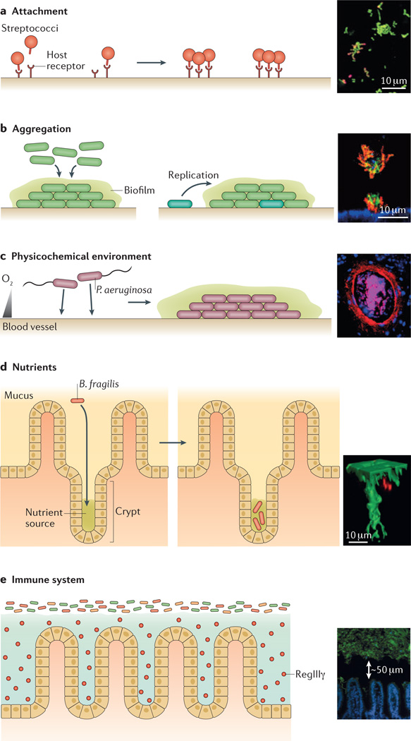
a | Attachment. Binding to specific host receptors can enable microorganisms to attach to surfaces. Therefore, the spatial distribution of such receptors may have an important role in determining the initial organization of microbial communities. For example, the insert shows streptococci (labelled in red; nonspecific nucleic acid stain labelled in green), which are initially sparse in human dental plaque communities, suggesting that the host receptors that streptococci adhere to are also sparse. b | Aggregation. Following attachment, microorganisms usually grow and organize into aggregates. Aggregation can involve collective behaviours (left panel) or can arise from single-cell founding events followed by clonal growth (right panel, in which the cyan shaded cell is the founding cell). For example, the insert shows Pseudomonas aeruginosa (labelled in red) forming microcolonies on the surface of epithelial cells (labelled in blue), in a process that is regulated by collective behaviour and depends on the expression of a type III secretion system (T3SS). The aggregate exhibits biofilm-like characteristics, such as staining positive for the matrix exopolysaccharide Psl (labelled in green). c | The physicochemical environment. Gradients, such as pH gradients and oxygen gradients, can influence the distribution of bacteria within communities. For example, the insert shows P. aeruginosa (labelled in red) concentrated around a vein (host cells are labelled in pink and blue) in a cross-section of a murine burn wound, in a process that may be regulated by oxygen availability. Scale bar not included. d | Nutrients. The ability to access and utilize certain nutrients in specific locations can also influence microbiogeography. For example, the insert shows Bacteroides fragilis (labelled in red), which carries a specific polysaccharide utilization locus that mediates its localization deep within an intestinal crypt (labelled in green). e | The immune system. Immune molecules, such as the lectin RegIIIγ, influence the distribution of microbial communities. For example, the insert shows how secretion of the lectin RegIIIγ by epithelial cells in the small intestine (labelled in blue) leads to the establishment of a 50-µm-wide gap between the intestinal surface and the microbiota (labelled in green). The insert in panel a is adapted from J. Bacteriol., 2003, 185, 3400–3409, doi:10.1128/JB.185.11.3400-3409.2003 and amended with permission from American Society for Microbiology. The insert in panel b is adapted from REF. . The insert in panel c is adapted from Infect. Immun., 2007, 75, 3715–3721, doi:10.1128/IAI.00586-07 and amended with permission from American Society for Microbiology. The insert in panel d is adapted from REF. , Nature Publishing Group. The insert in panel e is adapted from Vaishnava, S. et al. The antibacterial lectin RegIIIγ promotes the spatial segregation of microbiota and host in the intestine. Science
334, 255–258 (2011). Reprinted with permission from AAAS.
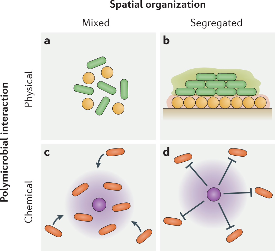
Chemical and physical polymicrobial interactions generate spatially mixed and segregated community patterns during infections. a | Co-aggregation, or intercellular binding, can cause spatial mixing. b | Excess production of biofilm polysaccharide (green and orange halos) can cause spatial segregation. c | Production of a beneficial metabolite (purple halo) can cause spatial mixing. d | Production of a harmful metabolite (purple halo) can cause spatial segregation.
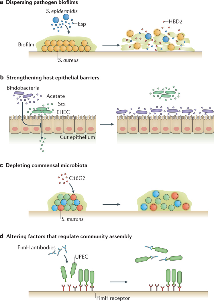
a | Dispersing pathogen biofilms. Spatially organizing into biofilms can make microorganisms resistant to challenges, such as host immune responses, so dispersing biofilms can provide a potential therapy against infections. For example, Staphylococcus epidermidis can destroy biofilms of Staphylococcus aureus through the production of the serine protease Esp. Furthermore, Esp enhances the susceptibility of S. aureus to human β-defensin 2 (HBD2), an antimicrobial peptide released during inflammation of the nasal cavity. b | Strengthening host epithelial barriers. Manipulating biogeography can also be used to prevent pathogens from crossing host barriers. For example, Shiga toxin (Stx) produced by enterohaemorrhagic Escherichia coli (EHEC) can cross the gut epithelium, but the introduction of commensal bifidobacteria can protect against fatal EHEC infection. Protective bifidobacteria can consume fructose and produce acetate, which induces an anti-inflammatory response in epithelial cells in the colon that lowers their susceptibility to Stx. c | Depleting commensal microbiota. Some commensal microorganisms can exacerbate infection by promoting pathogen virulence. Therefore, in situations in which targeting the pathogen directly has proven ineffective, depleting these commensals could represent an alternative therapeutic strategy. The feasibility of this approach is demonstrated by the synthetic antimicrobial peptide C16G2, which selectively kills Streptococcus mutans (in red), a commensal known to be involved in the progression of dental caries. Treating oral biofilms with C16G2 not only depletes communities of S. mutans (red cocci) but also other community members that directly interact with S. mutans. d | Altering factors that regulate community assembly. Masking microbial attachment sites, or reversing or eliminating the molecular gradients that give rise to virulence-associated spatial organizations, can eliminate microbiogeography. For example, a vaccine designed against the FimH adhesin, used by uropathogenic strains of E. coli (UPEC) to adhere to the bladder epithelium, is highly effective at limiting attachment and colonization of this pathogen in bladder infection models.
References
-
- Hall-Stoodley L, Costerton JW, Stoodley P. Bacterial biofilms: from the natural environment to infectious diseases. Nat. Rev. Microbiol. 2004;2:95–108. - PubMed
-
- Kaufmann SH, Schaible UE. 100th anniversary of Robert Koch’s Nobel Prize for the discovery of the tubercle bacillus. Trends Microbiol. 2005;13:469–475. - PubMed
-
- Smith H. The role of microbial interactions in infectious disease. Phil. Trans. R. Soc. Lond. B. Biol. Sci. 1982;297:551–561. - PubMed
Publication types
MeSH terms
Grants and funding
LinkOut - more resources
Full Text Sources
Other Literature Sources
