. 2015 Dec 30:12:247.
doi: 10.1186/s12974-015-0464-8.

Modulation of post-stroke degenerative and regenerative processes and subacute protection by site-targeted inhibition of the alternative pathway of complement

Affiliations

Modulation of post-stroke degenerative and regenerative processes and subacute protection by site-targeted inhibition of the alternative pathway of complement

Ali Alawieh et al. J Neuroinflammation. .

Abstract

Background: Complement promotes neuroinflammation and injury in models of stroke. However, complement is also being increasingly implicated in repair and regeneration after central nervous system (CNS) injury, and some complement deficiencies have been shown to provide acute, but not subacute, protection after murine stroke. Here, we investigate the dual role of complement in injury and repair after cerebral ischemia and reperfusion.

Methods: We used complement-deficient mice and different complement inhibitors in a model of transient middle cerebral artery occlusion to investigate complement-dependent cellular and molecular changes that occur through the subacute phase after stroke.

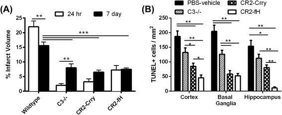
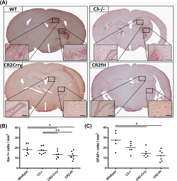
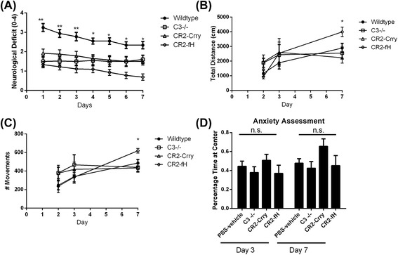
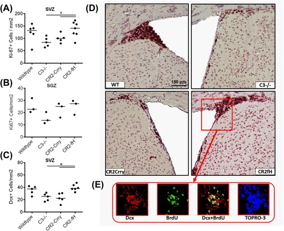
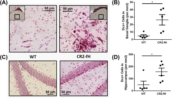
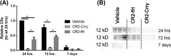
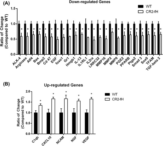
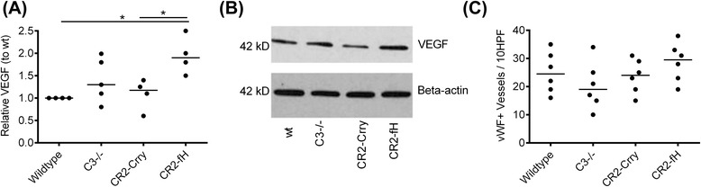
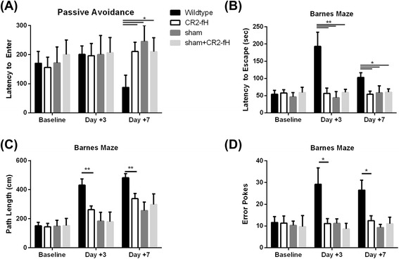
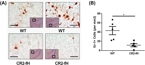
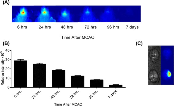
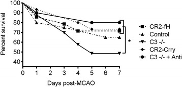
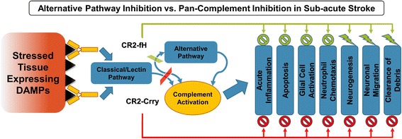
Results: C3 deficiency and site-targeted complement inhibition with either CR2-Crry (inhibits all pathways) or CR2-fH (inhibits alternative pathway) significantly reduced infarct size, reduced apoptotic cell death, and improved neurological deficit score in the acute phase after stroke. However, only in CR2-fH-treated mice was there sustained protection with no evolution of injury in the subacute phase. Whereas both inhibitors significantly reduced microglia/macrophage activation and astrogliosis in the subacute phase, only CR2-fH improved neurological deficit and locomotor function, maintained neurogenesis markers, enhanced neuronal migration, and increased VEGF expression. These findings in CR2-fH-treated mice correlated with improved performance in spatial learning and passive avoidance tasks. The complement anaphylatoxins have been implicated in repair and regenerative mechanisms after CNS injury, and in this context CR2-fH significantly reduced, but did not eliminate the generation of C5a within the brain, unlike CR2-Crry that completely blocked C5a generation. Gene expression profiling revealed that CR2-fH treatment downregulated genes associated with apoptosis, TGFβ signaling, and neutrophil activation, and decreased neutrophil infiltration was confirmed by immunohistochemistry. CR2-fH upregulated genes for neural growth factor and mediators of neurogenesis and neuronal migration. Live animal imaging demonstrated that following intravenous injection, CR2-fH targeted specifically to the post-ischemic brain, with a tissue half-life of 48.5 h. Finally, unlike C3 deficiency, targeted complement inhibition did not increase susceptibility to lethal post-stroke infection, an important consideration for stroke patients.

Conclusions: Ischemic brain tissue-targeted and selective inhibition of alternative complement pathway provide self-limiting inhibition of complement activation and reduces acute injury while maintaining complement-dependent recovery mechanisms into the subacute phase after stroke.


Figures

Fig. 1
Fig. 1
Complement inhibition reduces cerebral injury and cell death following MCAO and 7 days reperfusion. a Infarct volumes after 60 min MCAO and either 24-h or 7 days reperfusion. Mean +/− SEM, n = 8–12 mice. **p < 0.01, ***p < 0.001, #p < 0.001. b Cell death in different brain regions as analyzed by TUNEL immunostaining at 7 days after MCAO. Mean +/− SEM (TUNEL-positive cells per square millimeter), n = 5–6 mice. *p < 0.05
Fig. 2
Fig. 2
Complement inhibition reduces microglia/macrophage activation and astrogliosis following MCAO and 7 days reperfusion. a Representative images of activated microglia/macrophages as assessed by Iba-1 immunohistochemical detection across the different groups. Scale bar = 200 μm. b Quantification of Iba-1 immunohistochemical staining. Mean +/− SEM (positive cells per square millimeter), n = 6–8 mice. *p < 0.05, **p < 0.01. c Quantification of reactive astrocytes as assessed by GFAP immunohistochemical detection. Mean +/− SEM (positive cells per square millimeter), n = 5–7 mice. *p < 0.05
Fig. 3
Fig. 3
Effect of complement inhibition on behavioral deficit and locomotor function after MCAO. a Neurological deficit scores (0–4) within the first week after MCAO. Mean +/− SEM, n = 8–13. One-way ANOVA, **p < 0.05 (wild-type compared to all other groups), *p < 0.05 (wild-type compared to CR2-fH only). b Anxiety assessment across the different groups measured as percent time spent at the center of an open field. Mean +/− SEM. (One-way ANOVA, n.s. p > 0.05). c, d Locomotor activity measured as total distance moved (c) or number of movements (d), using an open field activity monitor. Determinations were made 2, 3, and 7 days after MCAO. Mean +/− SEM, n = 8–13. One-way ANOVA, *p < 0.05(CR2-fH-treated compared to wild-type)
Fig. 4
Fig. 4
CR2-fH increases subventricular zone neuronal proliferation and neuroblast presence at 7 days post-MCAO. a Doublecortin (Dcx+) neuroblasts within the ipsilateral SVZ at 7 days post-MCAO. Bar = Mean, n = 5–7. b Ki-67+ proliferating cells within the ipsilateral subventricular zone (SVZ) of the lateral ventricles. Bar = Mean. c, the subgranular zone (SGZ) of the dentate gyrus. Bar = Mean (density calculated from five random HPF), n = 3–7. *p < 0.05. d Representative images of immunostained sections, ×20 magnification. Scale bar = 150 μm. e Representative immunofluorescence double staining of SVG in CR2-fH-treated mice with anti-BrdU (green) and anti-Dcx (red), showing co-localization of signal
Fig. 5
Fig. 5
CR2-fH increases neuroblasts migration to perilesional basal ganglia and hippocampus at 7 days post-MCAO. a Representative images of neuroblast migration to the basal ganglia. b Quantification of number of Dcx + cells in basal ganglia. Bar = Mean +/− SEM, n = 5. *p < 0.05. c Representative images of neuroblasts in the hippocampus. d Quantification of number of Dcx + cells in hippocampus. Bar = Mean +/− SEM, n = 5–6. *p < 0.05
Fig. 6
Fig. 6
Different levels of C5a generation in brains from CR2-fH- and CR2-Crry-treated mice through subacute phase after stroke. a Relative C5a expression as determined by density scan of Western blot of ipsilateral brain tissue extract. Shown are relative expression levels normalized to level in wt control mice at 24 h. Mean +/− SEM, n = 4 animals per group, *p < 0.05. b Representative Western blot for C5a in brain homogenate
Fig. 7
Fig. 7
Nanostring analysis of changes in mRNA expression profile secondary to CR2-fH treatment at 7 days post-MCAO. a Genes with significantly reduced expression after CR2-fH treatment that exhibit more than 0.8-fold reduction in expression levels. Levels are normalized to the Mean of the wt controls. Mean +/− SEM, n = 4, *p < 0.05. b Genes with significantly increased expression after CR2-fH treatment that exhibit more than 1.25-fold reduction in expression levels. Levels are normalized to the Mean of the wt controls. Mean +/− SEM, n = 4, *p < 0.05. A complete dataset of different analyzed transcripts is provided in Additional file 1
Fig. 8
Fig. 8
CR2-fH increases relative VEGF expression but not angiogenesis in the ipsilateral brain at 7 days post-MCAO. a VEGF expression as determined by Western blot analysis of ipsilateral brain tissue extract. Shown are relative expression levels normalized to level in wt control mice, with β-actin serving as a loading control. Bar = Median, n = 4, *p < 0.05. b Representative Western blot for analysis of VEGF expression. c Von Willibrand factor positive (vWF+) vessels within the ipsilateral brain as detected by immunohistochemistry. Bar = Median (positive vessels per ten high-powered fields), n = 6
Fig. 9
Fig. 9
CR2-fH improves cognitive performance 7 days post-MCAO. a CR2-fH improves performance on passive avoidance task 7 days after MCAO. Shown is latency to enter a dark box associated with a shock. Mice were given a trial to associate a shock with the dark side of an apparatus, and latency to enter dark side was evaluated on days 3 and 7 post-MCAO. Mean +/− SEM, n = 7–10, *p < 0.05. bd, CR2-fH reduces spatial memory deficits in Barnes maze task in the subacute phase after MCAO. Mice were trained on the maze for 5 days before surgery and then tested on days 3 and 7 after MCAO for latency to escape (b), path length (c), and number of error pokes (d). Mean +/− SEM, n = 9–10, *p < 0.05, **p < 0.01
Fig. 10
Fig. 10
CR2-fH reduces brain neutrophil infiltration at 7 days after MCAO. a Representative images of neutrophil infiltration assessed by immunohistochemical staining with anti-Gr-1 antibody. Scale bar = 50 μm. b Quantification of Gr-1+ cells using 10 HPF/brain. Bar = Mean +/− SEM, n = 5, *p < 0.05
Fig. 11
Fig. 11
CR2-fH localizes to the brain after MCAO and specifically targets the ipsilateral lesion site. Fluorescently labeled CR2-fH was administered as described for therapeutic protocol, and localization of CR2-fH was visualized by in vivo fluorescence tomography. a Representative images of head scans of a single mouse taken at indicated times. b Quantification of fluorescent signal. Mean +/− SEM, n = 5 (24–72 h), n = 4 (72 h), n = 3 (7 days). c, Ex vivo image of brain removed 7 days after MCAO and injection of labeled CR2-fH (lower) or PBS (upper)
Fig. 12
Fig. 12
CR2-fH and CR2-Crry treatment but not C3 deficiency improves survival at 7 days post-MCAO. Kaplan-Meier survival analysis of C3-deficient or complement inhibitor-treated mice over a 7-day period post-MCAO. One group of C3-deficient mice received antibiotic prophylaxis (C3 −/− + Anti), n = 19–33, *p < 0.05
Fig. 13
Fig. 13
Schematic depicting complement activation and the effects of CR2-Crry vs. CR2-fH on complement-dependent effector mechanisms after ischemic stroke

References

    1. Komotar RJ, Kim GH, Otten ML, Hassid B, Mocco J, Sughrue ME, et al. The role of complement in stroke therapy. Adv Exp Med Biol. 2008;632:23–33. - PubMed
    1. Gorsuch WB, Chrysanthou E, Schwaeble WJ, Stahl GL. The complement system in ischemia-reperfusion injuries. Immunobiology. 2012;217:1026–1033. doi: 10.1016/j.imbio.2012.07.024. - DOI - PMC - PubMed
    1. Alawieh A, Elvington A, Tomlinson S. Complement in the homeostatic and ischemic brain. Front Immunol. 2015;6:417. doi: 10.3389/fimmu.2015.00417. - DOI - PMC - PubMed
    1. Elvington A, Atkinson C, Zhu H, Yu J, Takahashi K, Stahl GL, et al. The alternative complement pathway propagates inflammation and injury in murine ischemic stroke. J Immunol. 2012;189:4640–4647. doi: 10.4049/jimmunol.1201904. - DOI - PMC - PubMed
    1. Orsini F, Villa P, Parrella S, Zangari R, Zanier ER, Gesuete R, et al. Targeting mannose-binding lectin confers long-lasting protection with a surprisingly wide therapeutic window in cerebral ischemia. Circulation. 2012;126:1484–1494. doi: 10.1161/CIRCULATIONAHA.112.103051. - DOI - PMC - PubMed

Publication types

Substances