. 2015 Dec 29;6(3):559-71.
doi: 10.1534/g3.115.025841.

Novel Heterotypic Rox Sites for Combinatorial Dre Recombination Strategies

Affiliations

Novel Heterotypic Rox Sites for Combinatorial Dre Recombination Strategies

Katherine Chuang et al. G3 (Bethesda). .

Abstract

Site-specific recombinases (SSRs) such as Cre are widely used in gene targeting and genetic approaches for cell labeling and manipulation. They mediate DNA strand exchange between two DNA molecules at dedicated recognition sites. Precise understanding of the Cre recombination mechanism, including the role of individual base pairs in its loxP target site, guided the generation of mutant lox sites that specifically recombine with themselves but not with the wild type loxP. This has led to the development of a variety of combinatorial Cre-dependent genetic strategies, such as multicolor reporters, irreversible inversions, or recombination-mediated cassette exchange. Dre, a Cre-related phage integrase that recognizes roxP sites, does not cross-react with the Cre-loxP system, but has similar recombination efficiency. We have previously described intersectional genetic strategies combining Dre and Cre. We now report a mutagenesis screen aimed at identifying roxP base pairs critical for self-recognition. We describe several rox variant sites that are incompatible with roxP, but are able to efficiently recombine with themselves in either purified systems or bacterial and eukaryotic tissue culture systems. These newly identified rox sites are not recognized by Cre, thus enabling potential combinatorial strategies involving Cre, Dre, and target loci including multiple loxP and roxP variants.

Keywords: Cre recombination; Dre recombinase; gene targeting; site specific recombination.


Figures

Figure 1
Figure 1
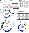
Random nucleotide libraries targeted at the roxP spacer region. (A) Comparison of roxP (top) and loxP (bottom) wild type sites. Horizontal arrows mark the inverted repeats for each target site and the black horizontal bars label identical base pairs. Regions highlighted in red correspond to the spacer region of the loxP site and the equivalent base pairs on the roxP site (numbered 1–8). The vertical arrowheads on the loxP sequence mark the sites of catalytic attack and strand break during Cre recombination. Mutations at positions 2 and 7 in the loxP spacer, highlighted in green, are used in the lox2272 mutant. (B) Libraries carrying random nucleotides indicated by N’s inserted at positions 3–6 (rox4R), 2–7 (rox6R) and 2, 3, 6, and 7 (rox2N) in the spacer region of roxP. (C) Plasmids and recombination test used in this paper. pGB2-Dre contains a replication cassette consisting of the repA protein and SC101 origin of replication (ensuring 1–5 copies/cell), a spectinomycin resistance gene (Spcr), and a Dre open reading frame driven by a lac promoter (LP). proxA-Kanr-roxB contains a pMB1 origin (generating >75 copies/cell), an ampicillin resistance gene, and a kanamycin (Kanr) resistance cassette, flanked by two rox sites arranged in direct orientation, roxA and roxB. If both rox sites are occupied by wild type roxP sequences (pPKanP vector), Dre recombination results in deletion of the intervening fragment containing the Kanr element. In the unrecombined plasmid, PvuI + PstI digestion results in 1.8 kb and 1.4 kb fragments, while after Dre recombination and Kanr removal the 1.8 kb fragment is reduced to 0.8 kb, while the 1.4 kb fragment (containing the pMB1-Ori) is unaffected. (D) Wild type roxP sites were inserted at the roxB position, while the mutant rox sites carrying the random nucleotide libraries shown in B were cloned into the roxA position, generating the p4RKanP, p6RKanP, and p2NKanP vectors.
Figure 2
Figure 2
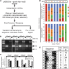
Screen strategy and results for identification of mutant rox sites. (A) Strategy used for recombination test (for p4RKanP, p6RKanP, or p2NKanP libraries) and for library screening (p2NKanP library). For details, see also Material and Methods. (B) PvuI + PstI restriction digest analysis of pooled target vectors before (−) and after (+) exposure to Dre recombination. Linearized pGB2-Dre can be seen in the (+) lanes (5.3 kb). Target vectors show the unrecombined restriction pattern (1.4 kb + 1.8 kb) before pGB2-Dre cotransformation (− lanes). After cotransformation (+ lanes), various degrees of recombination (0.8 kb band) can be seen. (C) Densitometric analysis of recombination efficiency of libraries and wild type control. (D) Nucleotide distributions at positions 1–8 in 82 individual p2NKanP minipreps sequenced after cotransformation with pGB2-Dre, and Kan selection (top) and 24 control p2NKanP minipreps from the unrecombined library (bottom). Original roxP sequence marked as wild type (WT) is indicated at the top. Positions 1, 4, 5, and 8 were not mutated in the rox2N spacer library. Numbers in the colored bars represent number of spacer sequences having the indicated nucleotide at the respective position. Nucleotide distributions were not significantly different between Dre-treated and untreated libraries at positions 3 (χ2 = 0.73, P = 0.87) and 6 (χ2 = 3.84, P = 0.28). However, after Dre selection, position 2 shows a highly significant bias toward G and C (χ2 = 18.44, P = 0.000357), and position 7 a significant bias toward G (χ2 = 9.99, P = 0.0186). (E) Most frequent spacers recovered from the screen. Nucleotides differing from consensus are shown in black. Occ. Nr. represents the number of times that specific sequence was observed. Clone number is the representative clone used for further studies.
Figure 3
Figure 3
Identified rox mutants are indeed unable to recombine with the wild type. Seven of the identified mutant spacers (A) were tested for the ability to recombine with the wild type roxP site, by cotransformation with pGB2-Dre in DH5α. Transformations were plated on ampicillin+ spectinomycin+, and two minipreps for each clone were amplified and diagnosed by restriction digest with PvuI + PstI (B, C). In addition, both clones were retransformed into DH5α plated on ampicillin plates, and replica plated using a velvet replicator onto kanamycin plates (D). (A) Schematic of the generic recombination target construct (top), and naming convention for the individual mutant constructs (bottom table). The roxB site is always roxP. The roxA site is either a wild type rox site (pPKanP) or one of the seven mutant spacers (Figure 2E). (B) Digest with PvuI + PstI. The 5.3 kb band represents the linearized pGB2-Dre. Both isolates of all seven mutant clones failed to recombine (1.8 + 1.4 kb bands), while the wild type isolates fully recombined (1.4 + 0.8 kb fragments). (C) Densitometric analysis confirms minimal recombination for mutants 7–9, and essentially no recombination for mutants 12–85. (D) Replica plating experiments for one of the two isolates for each clone, as well as the wild type control. For each clone, the number is indicated in the bottom left corner. Top rows are initial ampicillin plates, and bottom rows replicates onto kanamycin plates. The ratio of colonies surviving on kanamycin vs. ampicillin plates is shown as percent in A, bottom table. Images of full plates are provided in Figure S2.
Figure 4
Figure 4
Rox mutants rox7, rox8, rox12, rox61, and rox85 recombine with themselves in the presence of Dre but not Cre recombinase. (A) Rox sites with spacers identical to clones 7, 8, 12, 61, and 85 (Figure 2E) were cloned at both the roxA and roxB positions in direct orientation, generating the p7Kan7, p8Kan8, p12Kan12, p61Kan61, and p85Kan85 constructs. Each construct, as well as the pPKanP positive control, were then exposed to Dre recombinase by the same cotransformation protocol outlined in Figure 3. Two separate isolates for each clone were then tested by PvuI + PstI restriction digestion (B, C) and replica plating (Figure S3, A, B, C, and F). All rox sites tested showed complete recombination (1.4 + 0.8 kb fragments) when exposed to Dre recombinase, and essentially no surviving colonies when replica plated from ampicillin to kanamycin plates (Figure S3A, and table in A, right hand column). Quantitations of B are shown in C. To test the sensitivity of the wild type roxP and mutants to Cre recombination, pPKanP and p7Kan7, p8Kan8, p12Kan12, p61Kan61, and p85Kan85 mutants were transformed in the BS1365 strain, constitutively expressing Cre. Transformations were plated on kanamycin and ampicillin, and two individual colonies from each clone were amplified and analyzed by PvuI + PstI restriction digestion (D, E). In addition, DNA from the two colonies was retransformed in DH5α, plated on ampicillin, and colonies replica plated onto kanamycin plates using a velvet replicator (Figure S3B). All tested constructs show the 1.8 and 1.4 kb fragments characteristic for lack of recombination (D) and resistance to kanamycin in the replica platting assay (Figure S3B and table in A, right hand column). Note that the BS1365 strain carries the Cre recombinase on a F’ element that also contains a kanamycin resistance gene. Therefore, kanamycin does not select against rox target vector recombinants in the initial BS1365 cotransformation step.
Figure 5
Figure 5
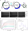
Self-recombination efficiencies of roxP and novel sites tested in purified system. (A) Diagram of in vitro recombination reaction. proxA-Kanr-roxB DNA is exposed to affinity-purified Dre, resulting in a recombined plasmid and a circle. Digestion with PvuI + PstI + MscI results in a common PstI-PvuI 1458 bp fragment spanning the pMB1 origin and part of the Ampr and two alternative, recombination-dependent sets of fragments. Without recombination, a MscI–PstI 713 bp fragment spanning the roxB site and a PvuI–MscI 1061 bp fragment spanning the roxA can be detected (see for instance the 0 min timepoint in B). After recombination, a PstI–PvuI fragment of 789 bp spanning the roxX site is observed in the remaining vector, and the 985 recombination circle is linearized by MscI. Without MscI digestion, the recombination circle migrates at about 600 bp (not shown). (B) Representative gels for the 0, 20, and 120 min time points reveal gradually accumulating recombination products. Lanes represent either marker (MW), or the prox-Kanr-rox vectors carrying either wild type (P) or spacers 7, 8, 12, 61, and 85 at both roxA and roxB sites (Figure 4A). Note that samples for the 20 and 120 min time points are taken from the same gel. (C) Densitometric analysis revealing percent recombination over the 120 min timecourse for wild type and all five mutant spacers. Solid lines represent medians, and dotted lines 25th and 75th quartiles for each set of samples (n = 6 data points/spacer and time point). (D) Box plots for the 60 min time point. Recombination efficiencies for each mutant spacer are shown as ratios to the wild type (P) from the same experiment (n = 10 data points/spacer). (Kolmogorov–Smirnov two sided test, significance levels ** P < 0.005, *** P < 0.001.)
Figure 6
Figure 6
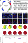
Rox mutants rox12 and rox85 recombine with themselves but not with roxP in eukaryotic cells. (A) In the prox-R-rox-G eukaryotic expression vector, a CMV promoter is driving transcription of mCherry before Dre recombination between the roxA and roxB sites, and of eGFP after recombination. rox12 and rox85 mutants or roxP wild type control were placed at both roxA and roxB sites to generate prox12-R-rox12-G, prox85-R-roxP-85 and proxP-R-roxP-G vectors, suitable for testing the ability of each rox site to recombine with itself. In addition, rox12 or rox85 mutants were cloned at the roxB site in combination with a roxP at the roxA site, to generate proxP-R-rox12-G and proxP-R-rox85-G, to test the compatibility between rox mutants and roxP wild type. (B, C) HEK293 cells stably expressing either the Dre (B) or Cre (C) recombinase were transfected with the constructs described in A. (D, E) Numbers in pie charts indicate the sums for red (mCherry only), green (eGFP only), and yellow (double positive) cells over six individual 20 × fields derived from two distinct transfected coverslips for each of the experimental conditions in B and C. No eGFP positive cells were seen in HEK293Dre cells transfected with proxP-R-rox12-G and proxP-R-rox85-G (B, D, columns 3 and 5) or HEK293Cre cells transfected with either one of the five constructs (C, E). (F) Box-whisker plots quantitating the percent of recombination positive cells in the HEK293Dre experiments. The percentage represents 100 × (eGFP+ + eGFP+mCherry+)/(eGFP+ + eGFP+mCherry+ + mCherry+). Statistical significance was determined by student t-test and Kolmogorov–Smirnov test (n.s. = P > 0.05, * = P < 0.05, ** = P < 0.005). Scale bar in C = 50 µm.
Figure 7
Figure 7
Efficient Dre recombination using an inversion–excision cassette (FREX) based on rox12 and roxP. (A) The pAAV-FREX-eGFP-mCherry eukaryotic viral expression vector contains a CAG promoter driving an expression cassette flanked by tandem rox12-roxP sites. The cassette contains eGFP and mCherry cDNAs placed in reverse orientation to each other, and separated by a triple repeat of a bidirectional SV40 transcription stop/polyadenylation signal. WPRE and bGH elements ensure mRNA stability and processing and AAV2 ITRs (not shown) allow for viral packaging. (B) Dre induces two alternative inversion reactions (double arrows) followed by irreversible excision (single arrow) reactions. The CAG promoter drives transcription of eGFP before, and of mCherry after, Dre recombination. HEK293-Dre (C) or HEK293-Cre (D) cells were transfected with FREX (column 1), prox-R-rox-G (column 2), or pAAVPTPY, a FLEX-based Cre reporter plasmid (column 3). (E, F) Numbers in pie charts for columns 1 and 2 indicate the sums for red (mCherry only), green (eGFP only), and yellow (double positive) cells. Results from cells transfected with pAAVPTPY (cyan) are shown in column 3 (E, F), comparing recombined cells to the total number of cells (DAPI, blue). Only Hek293Cre cells recombined the PTPY construct. (G) Box-whisker plots quantitating the percent of recombination positive cells. The percentage represents 100 × (mCherry+ + eGFP+mCherry+)/(eGFP+ + eGFP+mCherry+ + mCherry+) for FREX transfections or 100 × (eGFP+ + eGFP+mCherry+)/(eGFP+ + eGFP+mCherry+ + mCherry+) for pRoxP-R-roxP-G transfections. Recombination of PTPY is represented by 100 × Cyan+/DAPI+. Recombination efficiency was not statistically significant between the FREX and pRoxP-R-roxP-G in Dre-expressing cells. Statistical significance was determined with student t-test and Kolmogorov–Smirnov test (n.s. = P > 0.05, ** = P < 0.005). Scale bar in C = 50 μm.

Similar articles

Cited by

References

    1. Anastassiadis K., Fu J., Patsch C., Hu S., Weidlich S., et al. , 2009. Dre recombinase, like Cre, is a highly efficient site-specific recombinase in E. coli, mammalian cells and mice. Dis. Model. Mech. 2: 508–515. - PubMed
    1. Atasoy D., Aponte Y., Su H. H., Sternson S. M., 2008. A FLEX switch targets Channelrhodopsin-2 to multiple cell types for imaging and long-range circuit mapping. J. Neurosci. 28: 7025–7030. - PMC - PubMed
    1. Badea T. C., Cahill H., Ecker J., Hattar S., Nathans J., 2009. Distinct roles of transcription factors brn3a and brn3b in controlling the development, morphology, and function of retinal ganglion cells. Neuron 61: 852–864. - PMC - PubMed
    1. Birling M.-C., Gofflot F., Warot X., 2009. Site-specific recombinases for manipulation of the mouse genome. Methods Mol. Biol. 561: 245–263. - PubMed
    1. Bischof J., Basler K., 2008. Recombinases and their use in gene activation, gene inactivation, and transgenesis. Methods Mol. Biol. 420: 175–195. - PubMed

Publication types