Review
. 2016 Apr;7(2):187-98.
doi: 10.1007/s13244-015-0459-0. Epub 2015 Dec 29.

MRI for the preoperative evaluation of femoroacetabular impingement

Affiliations
Review

MRI for the preoperative evaluation of femoroacetabular impingement

Angela E Li et al. Insights Imaging. 2016 Apr.

Abstract

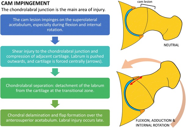
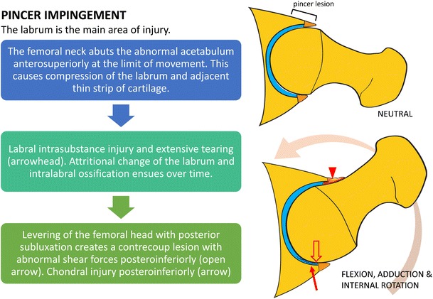
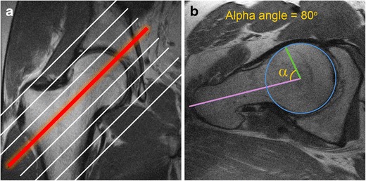
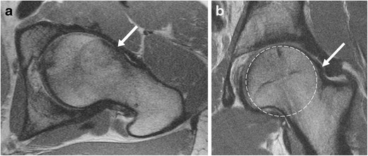
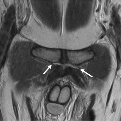
Femoroacetabular impingement (FAI) refers to a condition characterized by impingement of the femoral head-neck junction against the acetabular rim, often due to underlying osseous and/or soft tissue morphological abnormalities. It is a common cause of hip pain and limited range of motion in young and middle-aged adults. Hip preservation surgery aims to correct the morphological variants seen in FAI, thereby relieving pain and improving function, and potentially preventing early osteoarthritis. The purpose of this article is to review the mechanisms of chondral and labral injury in FAI to facilitate an understanding of patterns of chondrolabral injury seen on MRI. Preoperative MRI evaluation of FAI should include assessment of osseous morphologic abnormalities, labral tears, cartilage status, and other associated compensatory injuries of the pelvis. As advanced chondral wear is the major relative contraindication for hip preservation surgery, MRI is useful in the selection of patients likely to benefit from surgery. Teaching points • The most common anatomical osseous abnormalities predisposing to FAI include cam and pincer lesions. • Morphological abnormalities, labral lesions, and cartilage status should be assessed. • In cam impingement, chondral wear most commonly occurs anterosuperiorly.• Pre-existing advanced osteoarthritis is the strongest predictor of poor outcomes after FAI surgery. • Injury to muscles and tendons or other pelvic structures can coexist with FAI.

Keywords: Arthroscopy; Cartilage; Femoroacetabular impingement; Magnetic resonance imaging; Preoperative.


Figures

Fig. 1
Fig. 1
Diagram outlining the mechanisms of chondral and labral injury in cam impingement. The main focus of injury is the chondrolabral junction. There is relative sparing of the labrum until later in the disease process
Fig. 2
Fig. 2
Diagram outlining the mechanisms of chondral and labral injury in pincer impingement. The labrum is the main focus of damage. Chondral injury is initially limited to a relatively small strip of cartilage at the transition zone
Fig. 3
Fig. 3
Obtaining Swiss axial images and calculating the alpha angle. a The Swiss axial (axial oblique) images are obtained along the long axis of the femoral neck. b To calculate the alpha angle, the axial oblique image through the midportion of the femoral neck (red line) is chosen. A circle is drawn over the femoral head cortex (blue circle). A line is drawn through the long axis of the femoral neck at its narrowest point (purple line), through the center of the femoral head. Another line is drawn from the center of the femoral neck to the point where the femoral head–neck junction meets the circle (green line). The alpha angle is the angle between the two lines. An alpha angle of >55° is considered a risk factor for FAI
Fig. 4
Fig. 4
Oblique axial (a) and coronal (b) proton density (PD)-weighted MRI images in a 30-year-old man with a cam deformity. There is an osseous protuberance (arrows) at the femoral head–neck junction anterolaterally, with loss of offset of the femoral head–neck junction. The physeal scar extends lateral to the circular region of the femoral head (dashed circle) on the coronal image
Fig. 5
Fig. 5
An 18-year-old girl with pincer deformity. (a) On an axial oblique PD-weighted MRI image superiorly, the anterior rim of the acetabulum (arrow) is located lateral to the posterior rim (arrowhead), indicative of superior acetabular retroversion (blue line). Superior acetabular retroversion of 8° is shown in this example. (b) Corresponding radiograph demonstrates the "crossover" sign where the anterior acetabular wall projects (red line) lateral to the posterior acetabular wall (black dashed line) superiorly
Fig. 6
Fig. 6
A 26-year-old man with FAI. a Coronal PD-weighted image shows a cam lesion at the femoral head–neck junction (open arrow). There is chondrolabral separation, with a cleft between the labrum and cartilage (arrow). A paralabral cyst is also seen (black arrowhead). b Sagittal PD-weighted image shows chondral delamination near the transition zone anterosuperiorly (white arrowhead)
Fig. 7
Fig. 7
A 46-year-old woman with combined cam and pincer impingement. a Sagittal PD-weighted MRI demonstrates separation at the chondrolabral junction (black arrow). b Arthroscopic photo in the same patient demonstrates acetabular cartilage (A), labrum (L), and the transition zone between (dots). There is chondrolabral separation between 1 and 3 o'clock (white arrow) and chondral delamination (black arrowheads) adjacent to the transition zone
Fig. 8
Fig. 8
A 48-year-old man with combined cam and pincer impingement. a Coronal PD-weighted MRI demonstrates labral degeneration with intralabral ossification (white arrow). Separation is seen at the chondrolabral junction (black arrow). There is moderate to high-grade chondral wear over the superior femoral head and anterosuperior dome (arrowheads). b Axial oblique PD-weighted image shows a non-displaced tear of the anterior and anterosuperior labrum (arrow), with an associated intralabral cyst (arrowhead). c Arthroscopy image in the same patient demonstrates the labrum (L), acetabular (A), and femoral head (F) articular surfaces. An intrasubstance labral tear between 12 and 4 o'clock (white arrows), impaction erythema (asterisk), and chondral delamination at the transition zone (arrowhead) are seen. d There is moderate chondral wear (black arrows) over the femoral head, and normal cartilage is seen adjacent to this area (F)
Fig. 9
Fig. 9
A 36-year-old man with cam-type FAI and severe osteoarthritis. Coronal (a) and sagittal (b) PD-weighted MR images of the right hip demonstrate chronic degeneration of the labrum (arrowhead). Chondral loss with extensive bone-on-bone contact is seen over the superior femoral head (white arrow). Subcapital femoral neck osteophytes are also seen (black arrow). Reactive synovitis with effusion is demonstrated (asterisk)
Fig. 10
Fig. 10
Sagittal PD-weighted MRI of the hip in a 30-year-old woman demonstrates mild chondral hyperintensity over the anterosuperior acetabular dome (arrowhead), with corresponding prolongation of relaxation times on T2 mapping and T1rho images (white arrows)
Fig. 11
Fig. 11
Calculation of femoral version corrected for distal femoral rotation. On the straight axial image(s) of the hip/pelvis covering the femoral head and neck, a line is drawn between the center of the femoral head and center of the femoral neck at its narrowest point to calculate the uncorrected femoral anteversion angle (A; A is a negative value when the femur is retroverted). To correct for distal femoral rotation, another line is drawn along the posterior border of the femoral condyles to calculate the angle (B; B is a negative value when the knee is internally rotated). Femoral version = A − B. A positive value indicates femoral anteversion. A negative value indicates femoral retroversion. Normal femoral anteversion is approximately 12–13°
Fig. 12
Fig. 12
Compensatory injuries associated with FAI. Due to the altered biomechanics, there is increased strain on surrounding joints, tendons, and muscles, predisposing to injury. Preoperative MRI evaluation of FAI should include assessment of these structures
Fig. 13
Fig. 13
Coronal PD-weighted image of the pubic symphysis in a 34-year-old man with cam-type FAI and athletic pubalgia. There are linear fluid signal intensity clefts bilaterally, larger on the left, indicative of tears of the adductor longus tendon origins

References

    1. Nawabi DH, Bedi A, Tibor LM, Magennis E, Kelly BT. The demographic characteristics of high-level and recreational athletes undergoing hip arthroscopy for femoroacetabular impingement: a sports-specific analysis. Arthroscopy: J Arthroscopic Related Surg: Off Publ Arthroscopy Assoc North Am Int Arthroscopy Assoc. 2014;30(3):398–405. doi: 10.1016/j.arthro.2013.12.010. - DOI - PubMed
    1. Guevara CJ, Pietrobon R, Carothers JT, Olson SA, Vail TP. Comprehensive morphologic evaluation of the hip in patients with symptomatic labral tear. Clin Orthop Relat Res. 2006;453:277–285. doi: 10.1097/01.blo.0000246536.90371.12. - DOI - PubMed
    1. Beck M, Kalhor M, Leunig M, Ganz R. Hip morphology influences the pattern of damage to the acetabular cartilage: femoroacetabular impingement as a cause of early osteoarthritis of the hip. J Bone Joint Surg. British Vol. 2005;87(7):1012–1018. doi: 10.1302/0301-620X.87B7.15203. - DOI - PubMed
    1. Ganz R, Leunig M, Leunig-Ganz K, Harris WH. The etiology of osteoarthritis of the hip: an integrated mechanical concept. Clin Orthop Relat Res. 2008;466(2):264–272. doi: 10.1007/s11999-007-0060-z. - DOI - PMC - PubMed
    1. Ganz R, Parvizi J, Beck M, Leunig M, Notzli H, Siebenrock KA. Femoroacetabular impingement: a cause for osteoarthritis of the hip. Clin Orthop Relat Res. 2003;417:112–120. - PubMed

LinkOut - more resources