. 2016 Mar 2;17(5):415-20.
doi: 10.1002/cbic.201500601. Epub 2016 Feb 16.

Efficient Total Chemical Synthesis of (13) C=(18) O Isotopomers of Human Insulin for Isotope-Edited FTIR

Affiliations

Efficient Total Chemical Synthesis of (13) C=(18) O Isotopomers of Human Insulin for Isotope-Edited FTIR

Balamurugan Dhayalan et al. Chembiochem. .

Abstract

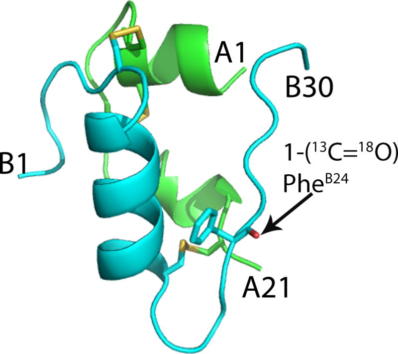
Isotope-edited two-dimensional Fourier transform infrared spectroscopy (2 D FTIR) can potentially provide a unique probe of protein structure and dynamics. However, general methods for the site-specific incorporation of stable (13) C=(18) O labels into the polypeptide backbone of the protein molecule have not yet been established. Here we describe, as a prototype for the incorporation of specific arrays of isotope labels, the total chemical synthesis-via a key ester insulin intermediate-of 97 % enriched [(1-(13) C=(18) O)Phe(B24) ] human insulin: stable-isotope labeled at a single backbone amide carbonyl. The amino acid sequence as well as the positions of the disulfide bonds and the correctly folded structure were unambiguously confirmed by the X-ray crystal structure of the synthetic protein molecule. In vitro assays of the isotope labeled [(1-(13) C=(18) O)Phe(B24) ] human insulin showed that it had full insulin receptor binding activity. Linear and 2 D IR spectra revealed a distinct red-shifted amide I carbonyl band peak at 1595 cm(-1) resulting from the (1-(13) C=(18) O)Phe(B24) backbone label. This work illustrates the utility of chemical synthesis to enable the application of advanced physical methods for the elucidation of the molecular basis of protein function.

Keywords: IR spectroscopy; chemical protein synthesis; human insulin; isotopic labeling; native chemical ligation.


Figures

Figure 1
Figure 1
Covalent structure of the human insulin protein molecule. The amino acid sequences of the 21 residue A chain and 30 residue B chain are given using the single letter code. Stable-isotope labeled residue PheB24 is highlighted in red.
Figure 2
Figure 2
[Upper panel] Stable-isotope labeled GlyA1-GluA4[OβThrB30-(1-13C=18O)PheB24-ThzB19]-CysA6-αthioester 1, R = CH2CO-Ala; [Lower panel] LCMS characterization of 1 after prep-HPLC purification (inset showing the online ESI-MS spectrum taken across the whole UV peak).): observed mass 2195.0 ± 0.2 Da; calculated mass, 2195.0 Da (monoisotopic) and 2195.4 Da (average isotopes).The chromatographic separations were performed on a C4 (2.1×50 mm) column using a linear gradient (5–45%) of solvent B in solvent A over 40 min (solvent A = 0.1% TFA in water, solvent B = 0.08% TFA in acetonitrile).
Figure 3
Figure 3
Folding stable-isotope labeled ester insulin, with concomitant formation of disulfide bonds. a) HPLC-traces showing the folding reaction at, t = 0 h and t = 2 h. Conditions: aqueous buffer containing 1.5 M GuHCl, 20 mM Tris, 8 mM Cysteine and 1 mM Cystine.HCl, pH 7.6, polypeptide concentration: 0.05 mg/mL; b) LC-MS data for the purified folded (1-13C=18O)PheB24 labeled human ester insulin (inset showing the online ESI-MS spectra taken across the whole of the main UV peak, and observed mass). The chromatographic separations were as described in Figure 2.
Figure 4
Figure 4
Saponification of isotope-labeled ester insulin to give [(1-13C=18O)PheB24]human insulin. Reaction was carried out in 25 mM LiOH in water at pH 12.2 and 4 °C. a) HPLC profiles showing the progress of the reaction as a function of time; b) LC-MS data for the [(1-13C=18O)PheB24]human insulin after HPLC purification (inset showing the online ESI-MS spectra across the whole of the UV absorbing peak). Reverse phase HPLC under the same conditions as for Figure 3. (Bottom panel) High resolution direct infusion ESI-MS spectra of [(1-13C=18O)PheB24]human insulin M+4H+ m/z region. Inset showing the monoisotope regions from [(1-13C=18O)PheB24]human insulin and [(1-13C=16O)PheB24]human insulin. The ratio between those two peaks was 96.7% which corresponds closely to the (1-13C=18O)PheB24 isotope enrichment in the final protein molecule.
Figure 5
Figure 5
X-ray crystal structure of [(1-13C=18O)PheB24]human insulin PDB code 5ENA. Cartoon representations of the A chain (green) and B chain (cyan). Disulfide bonds are shown in yellow. The site of isotope labeling at (1-13C=18O)PheB24 is shown as stick representation (O, red).
Figure 6
Figure 6
FTIR and 2D IR spectra of [(1-13C=18O)PheB24]human insulin in deuterated solvent at a concentration of 5 mg/mL in phosphate buffer and 20% ethanol at pD 2.0. (A) Linear FTIR spectra for the insulin amide I carbonyl stretch. [Inset: the FTIR difference spectrum of [(1-13C=18O)PheB24]human insulin from human insulin for the spectral region of the isotope label] (B) Temperature difference FTIR spectra of the (13C=18O)PheB24 isotope label relative to 55°C; 15° (blue) to 55°C (red). (C) 2D IR spectra at 15°C and (D) 55°C (waiting time of 150 fs).
Scheme 1
Scheme 1
Synthesis of [(1-13C=18O)PheB24]human insulin.
Scheme 2
Scheme 2
Preparation of the stable-isotope labeled segment GlyA1-GluA4[OβThrB30-(1-13C=18O)PheB24-ThzB19]-CysA6-αthioester 1 by solid phase peptide synthesis. Conditions: i) Boc-Ala-OCH2Ph-CH2COOH, DIC, DCM; ii) a) TFA, b) Trt-SCH2COOH, HBTU, DIEA, DMF; iii) a) TFA/TIPS/water (95:2.5:2.5 v/v), b) BocAA-OH, HBTU, DIEA, DMF (SPPS, 2 cycles); iv) a) TFA, b) Boc-Glu[Oβ(Alloc-L-Thr-α-OcHex)]-OH, HBTU, DIEA; v) a) TFA, BocAA-OH, HBTU, DIEA, DMF (SPPS, 3 cycles); b) TFA followed by 25% DIEA/DMF, 2-Cl-ZOSu, DMF; vi) a) Pd(PPh3)4, PhSiH3, DCM, b) BocAA-OH, HBTU, DIEA, DMF (SPPS, 11 cycles); HF-p-cresol.

References

    1. Le Sueur AL, Horness RE, Thielges MC. Analyst. 2015;140:4336–4349. - PMC - PubMed
    1. Baiz CR, Reppert M, Tokmakoff A. Ultrafast Infrared Vibrational Spectroscopy. CRC Press; 2013. pp. 361–404.
    1. Baiz CR, Tokmakoff A. Biophys J. 2015;108:1747–1757. - PMC - PubMed
    1. Courter JR, Abdo M, Brown SP, Tucker MJ, Hochstrasser RM, Smith AB. J Org Chem. 2014;79:759–768. - PMC - PubMed
    1. Ghosh A, Wang J, Moroz YS, Korendovych IV, Zanni M, DeGrado WF, Gai F, Hochstrasser RM. J Chem Phys. 2014;140:235105/1–235105/9. - PMC - PubMed

Publication types

MeSH terms