Review
. 2016 Feb;30(2):135-47.
doi: 10.1007/s40263-015-0297-0.

The Direct Effects of Fingolimod in the Central Nervous System: Implications for Relapsing Multiple Sclerosis

Affiliations
Review

The Direct Effects of Fingolimod in the Central Nervous System: Implications for Relapsing Multiple Sclerosis

Samuel F Hunter et al. CNS Drugs. 2016 Feb.

Abstract

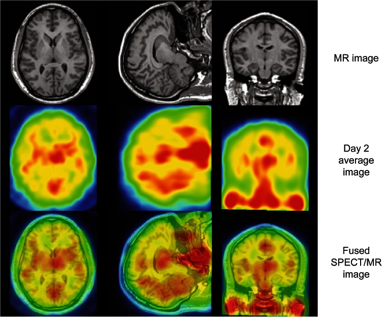
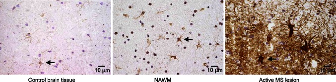
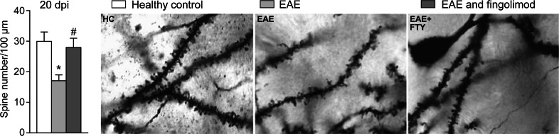
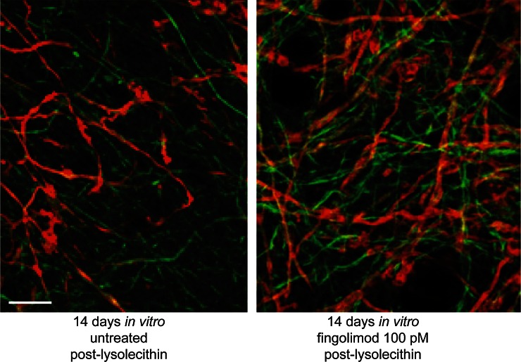
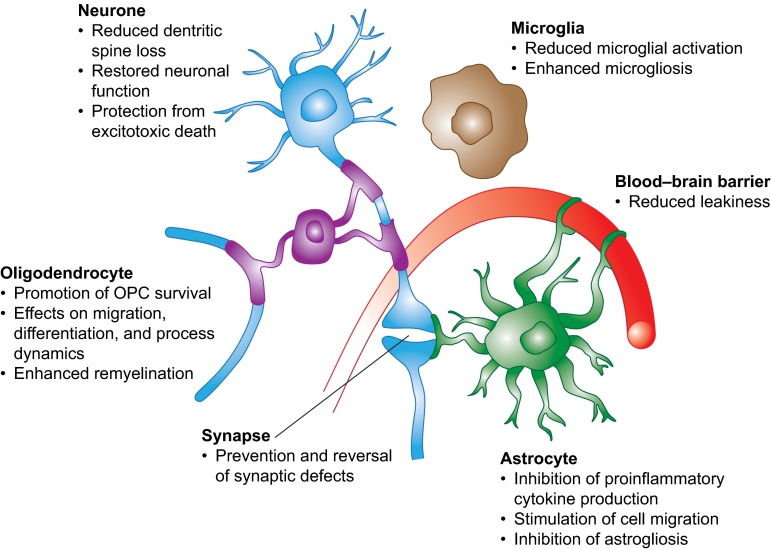
Fingolimod, a structural analog of sphingosine derived from fungal metabolites, is a functional antagonist of the G-protein-coupled sphingosine 1-phosphate (S1P) receptors S1P(1,3,4,5). In the treatment of relapsing forms of multiple sclerosis (RMS), fingolimod acts by reversibly retaining central memory T cells and naïve T cells in lymph nodes, thereby reducing the recirculation of autoreactive lymphocytes to the central nervous system (CNS). Fingolimod also has differential effects on the trafficking and function of B-cell subtypes and natural killer (NK) cells in peripheral blood and the CNS. Fingolimod also crosses the blood-brain barrier (BBB) and accumulates in the CNS. Experimental evidence increasingly supports a direct action of fingolimod within the CNS on brain cells, providing protection against the neurodegenerative component of RMS. We review the direct influence of this compound on CNS pathogenesis in RMS, including the central effects of fingolimod in animal models of MS and on neural cell types that express S1P receptors, such as astrocytes, BBB endothelial cells, microglia, neurones, and oligodendrocytes, which are all involved in RMS pathology.


Figures

Fig. 1
Fig. 1
Distribution of the fingolimod analog, iodine-123-labelled BZM055, in the human brain. MRI, mean SPECT image on day 2, and fused SPECT/MRI for a representative patient, depicting the brain uptake of the fingolimod analog [123I]BZM055. Chromatic changes indicate the range of no or low BZM055 uptake (blue/green), to moderate or high (yellow/red) uptake. Note that deep nuclei appear to exhibit the highest BZM055 uptake, however, there was no evidence of higher uptake in white matter relative to grey matter. Reproduced from Tamagnan et al. [55], with permission. MRI magnetic resonance imaging, SPECT single-photon emission computed tomography
Fig. 2
Fig. 2
Immunohistochemical staining for S1P1. Compared with control brain tissue, S1P1 staining on astrocytes was upregulated in NAWM in MS, and to the greatest extent in reactive astrocytes in active MS lesions. MS multiple sclerosis, NAWM normal-appearing white matter, S1P sphingosine 1-phosphate. Reproduced from Van Doorn et al. [68], with permission
Fig. 3
Fig. 3
Effect of fingolimod on dendritic spine loss in a mouse model of EAE. Prophylactic fingolimod treatment reduced the dendritic spine loss observed during the acute phase of EAE (expressed as spine density: number per 100 μm). Examples of single-section Golgi preparations from healthy control mice (n = 5), untreated EAE mice (n = 5), and prophylactic fingolimod-treated EAE mice (n = 5) at 20 dpi. dpi dots per inch, EAE experimental autoimmune encephalomyelitis, FTY fingolimod, HC healthy control. Reproduced from Rossi et al. [74], with permission. *p < 0.05 vs. control; # p < 0.05 vs. untreated EAE
Fig. 4
Fig. 4
Recovery of myelin in non-immune-mediated demyelinated cultures. Representative images of demyelinated slices, control or treated with fingolimod (100 pM) for 14 days in vitro post-lysolecithin, immunostained against myelin (MBP; red) and axons (NFM; green). Fingolimod increases the amount of myelin associated with axons compared with untreated control. Scale bar 20 µm. MBP myelin basic protein, NFM neurofilament. Reproduced from Miron et al. [71], with permission
Fig. 5
Fig. 5
Summary of the effects of fingolimod treatment on different cells in the central nervous system. OPC oligodendrocyte precursor cell. Reproduced from Groves et al. [60], with permission

References

    1. Brinkmann V. FTY720 (fingolimod) in multiple sclerosis: therapeutic effects in the immune and the central nervous system. Br J Pharmacol. 2009;158(5):1173–1182. doi: 10.1111/j.1476-5381.2009.00451.x. - DOI - PMC - PubMed
    1. Chun J, Hartung HP. Mechanism of action of oral fingolimod (FTY720) in multiple sclerosis. Clin Neuropharmacol. 2010;33(2):91–101. doi: 10.1097/WNF.0b013e3181cbf825. - DOI - PMC - PubMed
    1. Brinkmann V. Sphingosine 1-phosphate receptors in health and disease: mechanistic insights from gene deletion studies and reverse pharmacology. Pharmacol Ther. 2007;115(1):84–105. doi: 10.1016/j.pharmthera.2007.04.006. - DOI - PubMed
    1. Food and Drug Administration. Gilenya US prescribing information. Revised August 2015. 2010 [cited 29 September 2015]. Available from: http://www.accessdata.fda.gov/drugsatfda_docs/label/2015/022527s019lbl.pdf.
    1. European Medicines Agency. Gilenya EU summary of product characteristics, latest update of May 2015. 2011 [cited 16 June 2015]. Available from: http://www.ema.europa.eu/docs/en_GB/document_library/EPAR_-_Product_Info....

Publication types

MeSH terms

Substances

LinkOut - more resources