. 2016 Aug 1;33(15):1436-49.
doi: 10.1089/neu.2015.4186. Epub 2016 Mar 29.

Fatty Acid Binding Protein 5 Modulates Docosahexaenoic Acid-Induced Recovery in Rats Undergoing Spinal Cord Injury

Affiliations

Fatty Acid Binding Protein 5 Modulates Docosahexaenoic Acid-Induced Recovery in Rats Undergoing Spinal Cord Injury

Johnny D Figueroa et al. J Neurotrauma. .

Abstract

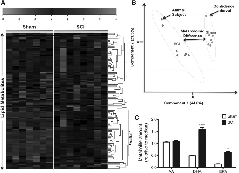
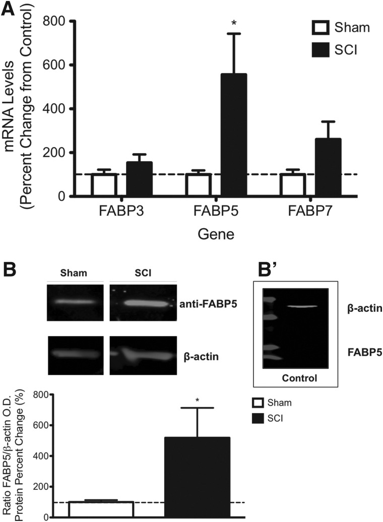
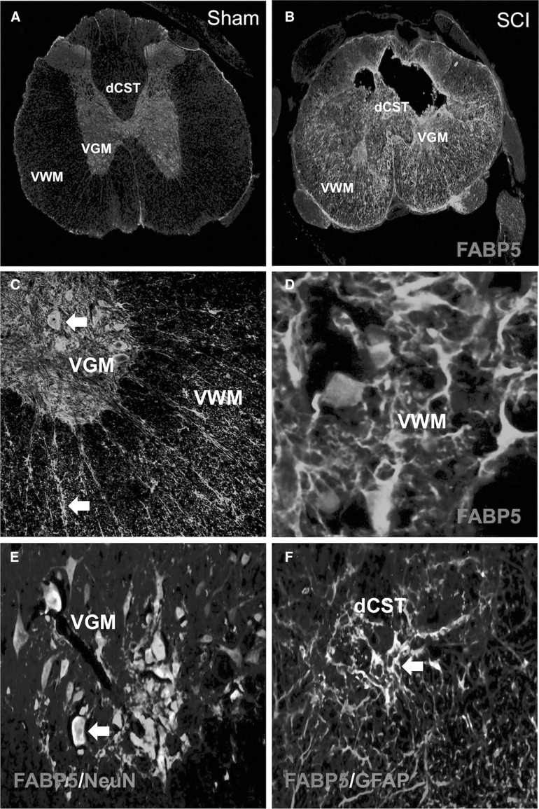
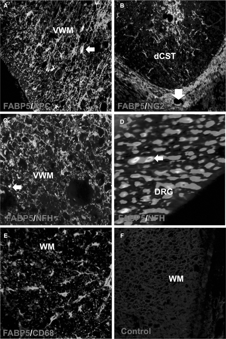
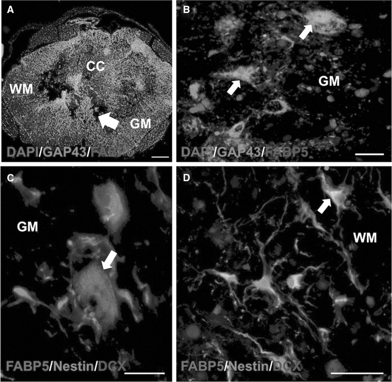
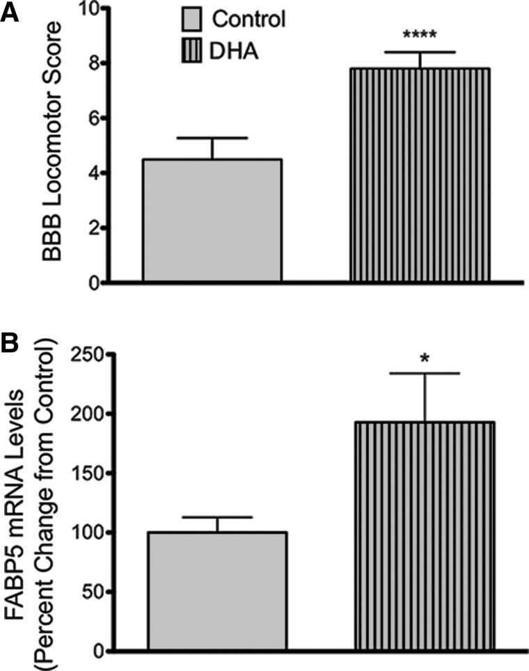
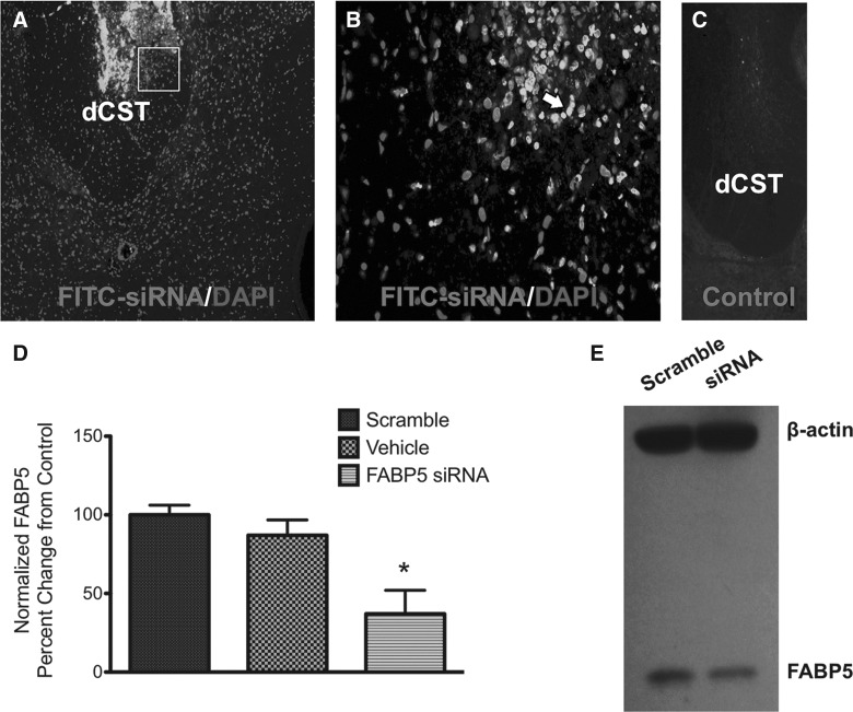
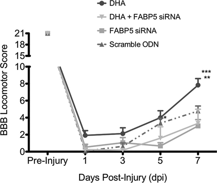
Omega-3 polyunsaturated fatty acids (n-3 PUFAs) promote functional recovery in rats undergoing spinal cord injury (SCI). However, the precise molecular mechanism coupling n-3 PUFAs to neurorestorative responses is not well understood. The aim of the present study was to determine the spatiotemporal expression of fatty acid binding protein 5 (FABP5) after contusive SCI and to investigate whether this protein plays a role in n-3 PUFA-mediated functional recovery post-SCI. We found that SCI resulted in a robust spinal cord up-regulation in FABP5 mRNA levels (556 ± 187%) and protein expression (518 ± 195%), when compared to sham-operated rats, at 7 days post-injury (dpi). This upregulation coincided with significant alterations in the metabolism of fatty acids in the injured spinal cord, as revealed by metabolomics-based lipid analyses. In particular, we found increased levels of the n-3 series PUFAs, particularly docosahexaenoic acid (DHA; 22:6 n-3) and eicosapentaenoic acid (EPA; 20:5 n-3) at 7 dpi. Animals consuming a diet rich in DHA and EPA exhibited a significant upregulation in FABP5 mRNA levels at 7 dpi. Immunofluorescence showed low basal FABP5 immunoreactivity in spinal cord ventral gray matter NeuN(+) neurons of sham-operated rats. SCI resulted in a robust induction of FABP5 in glial (GFAP(+), APC(+), and NG2(+)) and precursor cells (DCX(+), nestin(+)). We found that continuous intrathecal administration of FABP5 silencing with small interfering RNA (2 μg) impaired spontaneous open-field locomotion post-SCI. Further, FABP5 siRNA administration hindered the beneficial effects of DHA to ameliorate functional recovery at 7 dpi. Altogether, our findings suggest that FABP5 may be an important player in the promotion of cellular uptake, transport, and/or metabolism of DHA post-SCI. Given the beneficial roles of n-3 PUFAs in ameliorating functional recovery, we propose that FABP5 is an important contributor to basic repair mechanisms in the injured spinal cord.

Keywords: DHA; locomotion; n-3 fatty acids; repair; spinal cord injury.


Figures

<b>FIG. 1.</b>
FIG. 1.
SCI leads to robust alterations to the PUFA neurometabolome during the subacute injury phase. Heatmap representation of unsupervised hierarchical clustering for detected PUFAs (rows) on each sample type (columns) (A). Shades of gray represent metabolite decreases and increases, respectively, relative to the median metabolite levels. See scale. PLS-DA score plot containing two first components (B). Each plot mark corresponds to an observation (individual rat spinal cord sample). The confidence ellipses illustrate the 95% confidence regions. Principal component 1 (x-axis) shows distinctive spectra between sham and injury groups. SCI led to a significant increase in levels of both DHA and EPA at 7 dpi (n = at least 7 rats; p < 0.05) (C). No changes were observed in levels of AA when comparing sham versus injury groups at 7 dpi (p > 0.05). AA, arachidonic acid; DHA, docosahexaenoic acid; dpi, days post-injury; EPA, eicosapentaenoic acid; PLS-DA, partial least squares discriminant analyses; PUFAs, polyunsaturated fatty acids; SCI, spinal cord injury.
<b>FIG. 2.</b>
FIG. 2.
FABP5 expression is induced by contusive SCI. SCI significantly increased levels of FABP5 mRNA levels at 7 dpi (p < 0.01) (A). Densitometric analyses of Western blotting showed increased protein levels in SCI rats when compared to sham controls at 7 dpi (n = at least 6 rats; p < 0.05) (B). Pre-bleed serum controls acquired from rabbits used to generate the FABP5 antibody were used to validate the specificity of the immunoreaction (B'). dpi, days post-injury; FABP5, fatty acid binding protein 5; mRNA, messenger RNA; O.D., optical density; SCI, spinal cord injury.
<b>FIG. 3.</b>
FIG. 3.
FABP5 is mostly expressed by neurons in sham animals and in glia in the injured spinal cord. Basal levels of FABP5 expression were detected in the spinal cord gray matter (A). Increased FABP5 immunoreactivity was observed in spinal cord ventral white (VWM) and gray matter (VGM) at 7 dpi (B). Interestingly, this expression was more prominent in neuron- and glial-like cells (C and D). Confocal imaging definitively colocalizes FABP5 to neurons (NeuN+) (E) and activated astrocytes (GFAP+) (F); see arrows. Scale bars: 20 μm. dCST, dorsal corticospinal tract; dpi, days post-injury; FABP5, fatty acid binding protein 5; GFAP, glial fibrillary acidic protein; NeuN, neuronal nuclei.
<b>FIG. 4.</b>
FIG. 4.
FABP5 immunoreactivity is mostly observed in glial cells post-SCI. FABP5 immunoreactivity was detected in oligodendrocytes (A) and precursor cells expressing the NG2 proteoglycan (B). Immunofluorescence photomicrographs revealed FABP5 protein expression in axons and cytoskeletal structures of the VWM (C) and DRG (D). FABP5 was not expressed in macrophages/monocytes (CD68) (F). Pre-immunization serum confirmed the specificity of the immunoreaction (G). Scale bars: 20 μm. APC, allophycocyanin; dCST, dorsal corticospinal tract; DRG, dorsal root ganglion; FABP5, fatty acid binding protein 5; NFH, neurofilament heavy polypeptide; NG2, neuron-glial antigen 2; SCI, spinal cord injury; VWM, ventral white matter; WM, white matter.
<b>FIG. 5.</b>
FIG. 5.
FABP5 expression is prominent in sprouting fibers and neural stem cells post-SCI. Confocal microphotographs show that FABP5 protein colocalized with cells and structures that were positive to the growth-associated protein 43 (GAP43+) (A and B). Notably, we observed FABP5 expression in neurogenic niches post-SCI, as evidenced by colocalization of FABP5 to GM cells positive to doublecortin (DCX) (C) and WM cells immuoreactive to nestin (D). Scale bars: 20 μm. DAPI, 4′,6-diamidino-2-phenylindole; FABP5, fatty acid binding protein 5; GM, gray matter; SCI, spinal cord injury; WM, white matter.
<b>FIG. 6.</b>
FIG. 6.
FABP5 is markedly upregulated at 7 dpi in animals fed DHA-rich diets. Injured rats consuming diets rich in DHA for 8 weeks pre-injury and 7 days post-injury showed improved locomotor recovery when compared to animals receiving control diets at 7 dpi (n = at least 24 animals; p < 0.0001) (A). Animals with a DHA-enriched diet presented an increased in levels of FABP5 mRNA expression when compared to injured animals receiving control diets (n = at least 7 rats; p < 0.05) (B). BBB, Basso-Beattie-Bresnahan locomotor scale; DHA, docosahexaenoic acid; dpi, days post-injury; FABP5, fatty acid binding protein 5; mRNA, messenger RNA.
<b>FIG. 7.</b>
FIG. 7.
FABP5 siRNA penetrates injured spinal cord and blocks FABP5 mRNA and protein expression. Photomicrograph of spinal cord section from an animal receiving intrathecal FITC-tagged siRNA demonstrates penetration of oligonucleotides (A). Confocal microscopy revealed that most staining was localized in cells of the dorsal white matter in the CST area (B). Fluorescence was absent in the spinal cord sections from animals receiving non-FITC-tagged ODNs (C). qRT-PCR analysis of FABP5 siRNA-treated spinal cords showed a 60% reduction in FABP5 mRNA levels relative to animals treated with control scrambled sequence oligonucleotides (n = at least 3 animals per group; p < 0.05) (D). As recently reported, immunoblotting of cells treated with FABP5 siRNA showed reduced protein levels (E). CST, corticospinal tract; DAPI, 4′,6-diamidino-2-phenylindole; dCST, dorsal corticospinal tract; FABP5, fatty acid binding protein 5; FITC, fluorescein isothiocyanate; mRNA, messenger RNA; ODNs, oligodeoxynucleotides; qRT-PCR, quantitative real-time polymerase chain reaction; siRNA, small interfering RNA.
<b>FIG. 8.</b>
FIG. 8.
FABP5 siRNA administration hinders locomotor recovery post-SCI. BBB locomotor scores were obtained every other day for 1 week post-SCI. Administration of 2 μg of FABP5 siRNA showed a transient impairment in locomotor recovery at 5 dpi when compared to animals receiving scrambled sequence oligos (n = 8–9 animals per group; two-way ANOVA; p < 0.01). Notably, concomitant administration of DHA and FABP5 siRNA abolished the beneficial effects of DHA to accelerate locomotor recovery at 7 dpi (n = 3–6 animals per group; two-way ANOVA; p < 0.001). *p < 0.05 when comparing FABP5 siRNA versus control at 5 dpi; **p < 0.001 when comparing DHA versus control at 7 dpi; ***p < 0.0001 when comparing DHA versus DHA + FABP5 siRNA at 7 dpi. ANOVA, analysis of variance; BBB, Basso-Beattie-Bresnahan locomotor scale; DHA, docosahexaenoic acid; dpi, days post-injury; FABP5, fatty acid binding protein 5; FITC, fluorescein isothiocyanate; ODN, oligodeoxynucleotide; SCI, spinal cord injury; siRNA, small interfering RNA.

Similar articles

Cited by

References

    1. Hulsebosch C.E. (2002). Recent advances in pathophysiology and treatment of spinal cord injury. Adv. Physiol. Educ. 26, 238–255 - PubMed
    1. Hagg T., and Oudega M. (2006). Degenerative and spontaneous regenerative processes after spinal cord injury. J. Neurotrauma 23, 264–280 - PubMed
    1. Yip P.K., and Malaspina A. (2012). Spinal cord trauma and the molecular point of no return. Mol. Neurodegener. 7, 6. - PMC - PubMed
    1. Figueroa J.D., and De Leon M. (2014). Neurorestorative targets of dietary long-chain omega-3 fatty acids in neurological injury. Mol. Neurobiol. 50, 197–213 - PMC - PubMed
    1. Anderson D.K., and Hall E.D. (1993). Pathophysiology of spinal cord trauma. Ann. Emerg. Med. 22, 987–992 - PubMed