Classification of follicular lymphoma: the effect of computer aid on pathologists grading
- PMID: 26715518
- PMCID: PMC4696238
- DOI: 10.1186/s12911-015-0235-6
Classification of follicular lymphoma: the effect of computer aid on pathologists grading
Abstract
Background: Follicular lymphoma (FL) is one of the most common lymphoid malignancies in the western world. FL cases are stratified into three histological grades based on the average centroblast count per high power field (HPF). The centroblast count is performed manually by the pathologist using an optical microscope and hematoxylin and eosin (H&E) stained tissue section. Although this is the current clinical practice, it suffers from high inter- and intra-observer variability and is vulnerable to sampling bias.
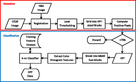
Methods: In this paper, we present a system, called Follicular Lymphoma Grading System (FLAGS), to assist the pathologist in grading FL cases. We also assess the effect of FLAGS on accuracy of expert and inexperienced readers. FLAGS automatically identifies possible HPFs for examination by analyzing H&E and CD20 stains, before classifying them into low or high risk categories. The pathologist is first asked to review the slides according to the current routine clinical practice, before being presented with FLAGS classification via color-coded map. The accuracy of the readers with and without FLAGS assistance is measured.
Results: FLAGS was used by four experts (board-certified hematopathologists) and seven pathology residents on 20 FL slides. Access to FLAGS improved overall reader accuracy with the biggest improvement seen among residents. An average AUC value of 0.75 was observed which generally indicates "acceptable" diagnostic performance.
Conclusions: The results of this study show that FLAGS can be useful in increasing the pathologists' accuracy in grading the tissue. To the best of our knowledge, this study measure, for the first time, the effect of computerized image analysis on pathologists' grading of follicular lymphoma. When fully developed, such systems have the potential to reduce sampling bias by examining an increased proportion of HPFs within follicle regions, as well as to reduce inter- and intra-reader variability.
Figures


Similar articles
-
Computer-aided detection of centroblasts for follicular lymphoma grading using adaptive likelihood-based cell segmentation.IEEE Trans Biomed Eng. 2010 Oct;57(10):2613-6. doi: 10.1109/TBME.2010.2055058. Epub 2010 Jun 28. IEEE Trans Biomed Eng. 2010. PMID: 20595077 Free PMC article.
-
A study of criteria for grading follicular lymphoma using a cell type classifier from pathology images based on complementary-label learning.Micron. 2024 Sep;184:103663. doi: 10.1016/j.micron.2024.103663. Epub 2024 May 30. Micron. 2024. PMID: 38843576
-
Computer-assisted quantification of CD3+ T cells in follicular lymphoma.Cytometry A. 2017 Jun;91(6):609-621. doi: 10.1002/cyto.a.23049. Epub 2017 Jan 22. Cytometry A. 2017. PMID: 28110507 Free PMC article.
-
Informatics Approaches to Address New Challenges in the Classification of Lymphoid Malignancies.JCO Clin Cancer Inform. 2018;2:CCI.17.00039. doi: 10.1200/CCI.17.00039. Epub 2018 Feb 9. JCO Clin Cancer Inform. 2018. PMID: 30637363 Free PMC article. Review.
-
Follicular lymphoma grade 3: review and updates.Clin Lymphoma Myeloma Leuk. 2014 Dec;14(6):431-5. doi: 10.1016/j.clml.2014.04.008. Epub 2014 Jun 12. Clin Lymphoma Myeloma Leuk. 2014. PMID: 25066038 Review.
Cited by
-
FOXP3-stained image analysis for follicular lymphoma: Optimal adaptive thresholding with maximal nucleus coverage.Proc SPIE Int Soc Opt Eng. 2017 Feb 11;10140:101400E. doi: 10.1117/12.2255671. Epub 2017 Mar 1. Proc SPIE Int Soc Opt Eng. 2017. PMID: 28579665 Free PMC article.
-
Role of artificial intelligence in haematolymphoid diagnostics.Histopathology. 2025 Jan;86(1):58-68. doi: 10.1111/his.15327. Epub 2024 Oct 22. Histopathology. 2025. PMID: 39435690 Free PMC article. Review.
-
Automated scoring methods for quantitative interpretation of Tumour infiltrating lymphocytes (TILs) in breast cancer: a systematic review.BMC Cancer. 2024 Sep 30;24(1):1202. doi: 10.1186/s12885-024-12962-8. BMC Cancer. 2024. PMID: 39350098 Free PMC article.
-
Primary follicular non-Hodgkin's lymphoma of the ureter: A case report and literature review.Oncol Lett. 2016 Jun;11(6):3939-3942. doi: 10.3892/ol.2016.4524. Epub 2016 May 5. Oncol Lett. 2016. PMID: 27313721 Free PMC article.
-
Detection of centroblast cells in H&E stained whole slide image based on object detection.Front Med (Lausanne). 2024 Feb 7;11:1303982. doi: 10.3389/fmed.2024.1303982. eCollection 2024. Front Med (Lausanne). 2024. PMID: 38384407 Free PMC article.
References
-
- Jaffe ES, Harris NL, Stein H, Vardiman JW. Tumours of haematopoietic and lymphoid tissues. Lyon, France: IRAC Press; 2008.
-
- Metter GE, Nathwani BN, Burke JS, Winberg CD, Mann RB, Barcos M, et al. Morphological sub-classification of follicular lymphoma: variability of diagnoses among hematopathologists, a collaborative study between the repository center and pathology panel for lymphoma clinical studies. J Clin Oncol. 1985;3:25–38. - PubMed
-
- Dick F, Van Lier S, Banks P, Frizzera G, Witrak G, Gibson R, et al. Use of the working formulation for non-Hodgkin’s lymphoma in epidemiological studies: agreement between reported diagnoses and a panel of experienced pathologists. J Natl Cancer Inst. 1987;78:1137–44. - PubMed
-
- The Non-Hodgkin Lymphoma Classification Project A clinical evaluation of the International Lymphoma Study Group classification of non-Hodgkin lymphoma. Blood. 1997;89:3909–3918. - PubMed
Publication types
MeSH terms
Grants and funding
LinkOut - more resources
Full Text Sources
Other Literature Sources

